Alveolar restoration following rapid maxillary expansion with and without corticotomy: A microcomputed tomography study in sheep
- Affiliations
-
- 1Department of Pediatric Dentistry and Orthodontics, Faculty of Dentistry, University of Malaya, Kuala Lumpur, Malaysia. myorth@yahoo.com
- 2Department of Restorative Dentistry, Wellness Research Cluster, University of Malaya, Kuala Lumpur, Malaysia.
- 3Department of Oro-maxillofacial Surgical and Medical Sciences, Faculty of Dentistry, University of Malaya, Kuala Lumpur, Malaysia.
- 4Department of Medical Imaging, Faculty of Health Sciences, Universiti Teknologi MARA (UiTM), Selangor, Malaysia.
- KMID: 2453060
- DOI: http://doi.org/10.4041/kjod.2019.49.4.235
Abstract
OBJECTIVE
This study examined bone microstructure restoration after rapid maxillary expansion (RME) with and without corticotomy over multiple retention periods.
METHODS
Eighteen male Dorper sheep were randomly distributed into three groups (n = 6 each group): group 1, RME with corticotomy on the buccal and palatal sides; group 2, conventional RME treatment; and group 3, no treatment. Post-RME, trabecular bone microstructure and new bone formation were evaluated by using microcomputed tomography (microCT) and histomorphometry after a 4- or 12-week retention period. Intergroup differences in bone quality and bone remodeling were analyzed by using two-way analysis of variance with Bonferroni post-hoc test.
RESULTS
The bone volume fraction (bone volume [BV]/total volume [TV]) values relative to the control in groups 1 and 2 were 54.40% to 69.88% after the 4-week retention period and returned to approximately 80% after the 12-week retention period. The pooled BV/TV values of the banded teeth in groups 1 and 2 were significantly lower than those of the control after the 4-week retention period (p < 0.05). However, after the 12-week retention period, the pooled BV/TV values in group 2 were significantly lower than those in groups 1 and 3 (p < 0.05). Histomorphological analysis showed that the new bone formation area in group 1 was approximately two to three times of those in group 2 and control.
CONCLUSIONS
Corticotomy significantly enhanced the restoration of bone quality after the retention periods for banded teeth. This benefit might result from the increased new bone formation after corticotomy.
MeSH Terms
Figure
-
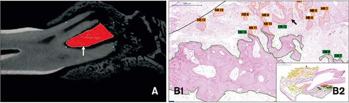
Figure 1 Region of interest (ROI) in quantification of bone microstructure (A) and new bone (NB) and old bone (OB) formation (B1 and B2). A, The furcation enclosed by the buccal and lingual roots of premolars was defined as the ROI (arrow) for measurements of bone microstructure. B, Magnification at hematoxylin and eosin (H&E) stain (×200) showing NB and OB. NB is defined as the amorphous eosinophilic material with porous bone and pale cement lines (black arrow). OB is the bone with a compact feature and prominent cement lines (white arrow) (B1); the total tissue area (outlined by the blue line) at H&E stain (×20) (B2).
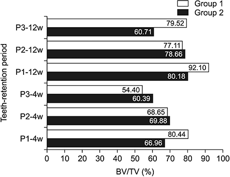
Figure 2 Relative values of bone volume fraction (BV/TV) at the premolar regions compared to the control values in groups 1 (corticotomy + rapid maxillary expansion [RME]) and 2 (conventional RME) after 4 and 12 weeks of retention. P1, First premolar; P2, second premolar; P3, third premolar; 4w, 4-week retention; 12w, 12-week retention.
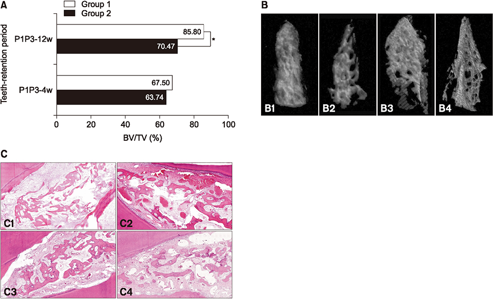
Figure 3 A, Relative values of bone volume fraction (BV/TV) values compared to the control values for the banded teeth (the first and third premolars) in groups 1 (corticotomy + rapid maxillary expansion [RME]) and 2 (conventional RME) after 4 and 12 weeks of retention. B, Alveolar trabecular bone microstructural images of group 1 after 4 (B1) and 12 weeks of retention (B3) and of group 2 after 4 (B2) and 12 weeks of retention (B4). C, Histological images at H&E stain (×100) showed that the bone volume in group 1 after 4 and 12 weeks of retention (C1 and C3) were higher than those in group 2 after 4 and 12 weeks of retention (C2 and C4) (H&E stain, ×100). P1, First premolar; P3, third premolar; 4w, 4-week retention; 12w, 12-week retention. *p < 0.05.
Reference
-
1. King EW. Relapse of orthodontic treatment. Angle Orthod. 1974; 44:300–315.2. Little RM, Dr. Robert M. Little on the University of Washington post-retention studies. J Clin Orthod. 2009; 43:723–727.3. Little RM. Clinical implications of the University of Washington post-retention studies. J Clin Orthod. 2009; 43:645–651.4. Gurel HG, Memili B, Erkan M, Sukurica Y. Long-term effects of rapid maxillary expansion followed by fixed appliances. Angle Orthod. 2010; 80:5–9.
Article5. Suri L, Taneja P. Surgically assisted rapid palatal expansion: a literature review. Am J Orthod Dentofacial Orthop. 2008; 133:290–302.
Article6. Johnston CD, Littlewood SJ. Retention in orthodontics. Br Dent J. 2015; 218:119–122.
Article7. van Leeuwen EJ, Maltha JC, Kuijpers-Jagtman AM, van't Hof MA. The effect of retention on orthodontic relapse after the use of small continuous or discontinuous forces. An experimental study in beagle dogs. Eur J Oral Sci. 2003; 111:111–116.
Article8. Godoy F, Godoy-Bezerra J, Rosenblatt A. Treatment of posterior crossbite comparing 2 appliances: a community-based trial. Am J Orthod Dentofacial Orthop. 2011; 139:45–52.
Article9. Cozzani M, Guiducci A, Mirenghi S, Mutinelli S, Siciliani G. Arch width changes with a rapid maxillary expansion appliance anchored to the primary teeth. Angle Orthod. 2007; 77:296–302.
Article10. Bell RA, LeCompte EJ. The effects of maxillary expansion using a quad-helix appliance during the deciduous and mixed dentitions. Am J Orthod. 1981; 79:152–161.
Article11. Petrén S, Bjerklin K, Bondemark L. Stability of unilateral posterior crossbite correction in the mixed dentition: a randomized clinical trial with a 3-year follow-up. Am J Orthod Dentofacial Orthop. 2011; 139:e73–e81.
Article12. Kole H. Surgical operations on the alveolar ridge to correct occlusal abnormalities. Oral Surg Oral Med Oral Pathol. 1959; 12:515–529.
Article13. Wilcko MT, Wilcko WM, Pulver JJ, Bissada NF, Bouquot JE. Accelerated osteogenic orthodontics technique: a 1-stage surgically facilitated rapid orthodontic technique with alveolar augmentation. J Oral Maxillofac Surg. 2009; 67:2149–2159.
Article14. Chung KR, Kim SH, Lee BS. Speedy surgical-orthodontic treatment with temporary anchorage devices as an alternative to orthognathic surgery. Am J Orthod Dentofacial Orthop. 2009; 135:787–798.
Article15. Lines PA. Adult rapid maxillary expansion with corticotomy. Am J Orthod. 1975; 67:44–56.
Article16. Echchadi ME, Benchikh B, Bellamine M, Kim SH. Corticotomy-assisted rapid maxillary expansion: a novel approach with a 3-year follow-up. Am J Orthod Dentofacial Orthop. 2015; 148:138–153.
Article17. Le MHT, Lau SF, Ibrahim N, Noor Hayaty AK, Radzi ZB. Adjunctive buccal and palatal corticotomy for adult maxillary expansion in an animal model. Korean J Orthod. 2018; 48:98–106.
Article18. Charan J, Kantharia ND. How to calculate sample size in animal studies? J Pharmacol Pharmacother. 2013; 4:303–306.
Article19. Ferguson DJ, Wilcko MT. Tooth movement mechanobiology: toward a unifying concept. In : Shroff B, editor. Biology of orthodontic tooth movement. Basel: Springer;2016. p. 13–44.20. Verna C, Zaffe D, Siciliani G. Histomorphometric study of bone reactions during orthodontic tooth movement in rats. Bone. 1999; 24:371–379.
Article21. Chang HW, Huang HL, Yu JH, Hsu JT, Li YF, Wu YF. Effects of orthodontic tooth movement on alveolar bone density. Clin Oral Investig. 2012; 16:679–688.
Article22. Hsu JT, Chang HW, Huang HL, Yu JH, Li YF, Tu MG. Bone density changes around teeth during orthodontic treatment. Clin Oral Investig. 2011; 15:511–519.
Article23. Panmekiate S, Ngonphloy N, Charoenkarn T, Faruangsaeng T, Pauwels R. Comparison of mandibular bone microarchitecture between micro-CT and CBCT images. Dentomaxillofac Radiol. 2015; 44:20140322.
Article24. Franzen TJ, Monjo M, Rubert M, Vandevska-Radunovic V. Expression of bone markers and micro-CT analysis of alveolar bone during orthodontic relapse. Orthod Craniofac Res. 2014; 17:249–258.
Article25. Martins DC, Souki BQ, Cheib PL, Silva GA, Reis ID, Oliveira DD, et al. Rapid maxillary expansion: do banded teeth develop more external root resorption than non-banded anchorage teeth? Angle Orthod. 2016; 86:39–45.
Article26. Langford SR. Root resorption extremes resulting from clinical RME. Am J Orthod. 1982; 81:371–377.
Article27. Sebaoun JD, Kantarci A, Turner JW, Carvalho RS, Van Dyke TE, Ferguson DJ. Modeling of trabecular bone and lamina dura following selective alveolar decortication in rats. J Periodontol. 2008; 79:1679–1688.
Article28. Bogoch E, Gschwend N, Rahn B, Moran E, Perren S. Healing of cancellous bone osteotomy in rabbits--part II: local reversal of arthritis-induced osteopenia after osteotomy. J Orthop Res. 1993; 11:292–298.
Article29. McBride MD, Campbell PM, Opperman LA, Dechow PC, Buschang PH. How does the amount of surgical insult affect bone around moving teeth? Am J Orthod Dentofacial Orthop. 2014; 145:S92–S99.
Article
- Full Text Links
-
- Actions
-
Cited
- CITED
-
- Close
- Share
-
- Similar articles
-
- Adjunctive buccal and palatal corticotomy for adult maxillary expansion in an animal model
- A posteroanterior cephalometric study on the change of maxilla by rapid palatal expansion
- Skeletal and dentoalveolar effects of different types of microimplant-assisted rapid palatal expansion
- A cone-beam computed tomography evaluation of buccal bone thickness following maxillary expansion
- A study on the effect of rapid maxillary expansion and its relapse
