Relationship between the maxillofacial skeletal pattern and the morphology of the mandibular symphysis: Structural equation modeling
- Affiliations
-
- 1Department of Orthodontics, Dental Research Institute, Pusan National University Dental Hospital, Yangsan, Korea. kimyongil@pusan.ac.kr
- 2Department of Management Information Systems, College of Business, Dong-A University, Busan, Korea.
- 3Department of Orthodontics, School of Dentistry, Showa University, Tokyo, Japan.
- 4Department of Orthodontics, Chang Gung Memorial Hospital, Kaohsiung, Taiwan.
- 5Department of Orthodontics, School of Dentistry, University of North Carolina at Chapel Hill, NC, USA.
- 6Institute of Translational Dental Sciences, School of Dentistry, Pusan National University, Busan, Korea.
- KMID: 2450226
- DOI: http://doi.org/10.4041/kjod.2019.49.3.170
Abstract
OBJECTIVE
The purpose of this study was to investigate the relationship between the facial skeletal patterns and the shape of the mandibular symphysis in adults with malocclusion by using a structural equation model (SEM).
METHODS
Ninety adults who had malocclusion and had records of facial skeletal measurements performed using cone-beam computed tomography were selected for this study. The skeletal measurements were classified into three groups (vertical, anteroposterior, and transverse). Cross-sectional images of the mandibular symphysis were analyzed using generalized Procrustes and principal component (PC) analyses. A SEM was constructed after the factors were extracted via factor analysis.
RESULTS
Two factors were extracted from the transverse, vertical, and anteroposterior skeletal measurements. Latent variables were extracted for each factor. PC1, PC2, and PC3 were selected to analyze the variations of the mandibular symphyseal shape. The SEM was constructed using the skeletal variables, PCs, and latent variables. The SEM showed that the vertical latent variable exerted the most influence on the mandibular symphyseal shape.
CONCLUSIONS
The relationship between the skeletal pattern and the mandibular symphysis was analyzed using a SEM, which showed that the vertical facial skeletal pattern had the highest effect on the shape of the mandibular symphysis.
Figure
-
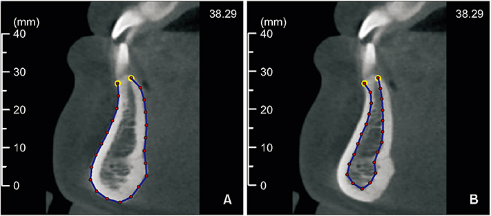
Figure 1 Obtaining the landmarks for the mandibular symphysis cross-sectional images. The outline of the mandibular symphysis is considered for the external (A) and internal (B) cortices. Two landmarks (yellow) and 21 semi-landmarks (red) are positioned along the outline of the mandibular cortex.
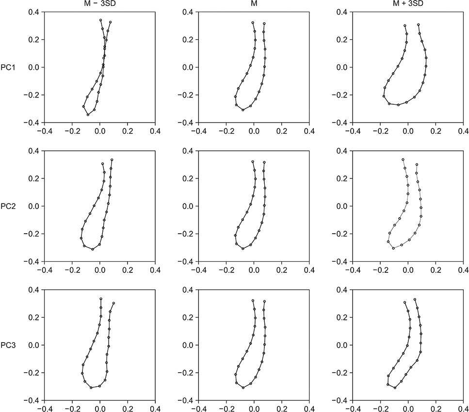
Figure 2 Principal component (PC) analysis of the internal cortices of the mandibular symphysis. M, Mean; SD, standard deviation.
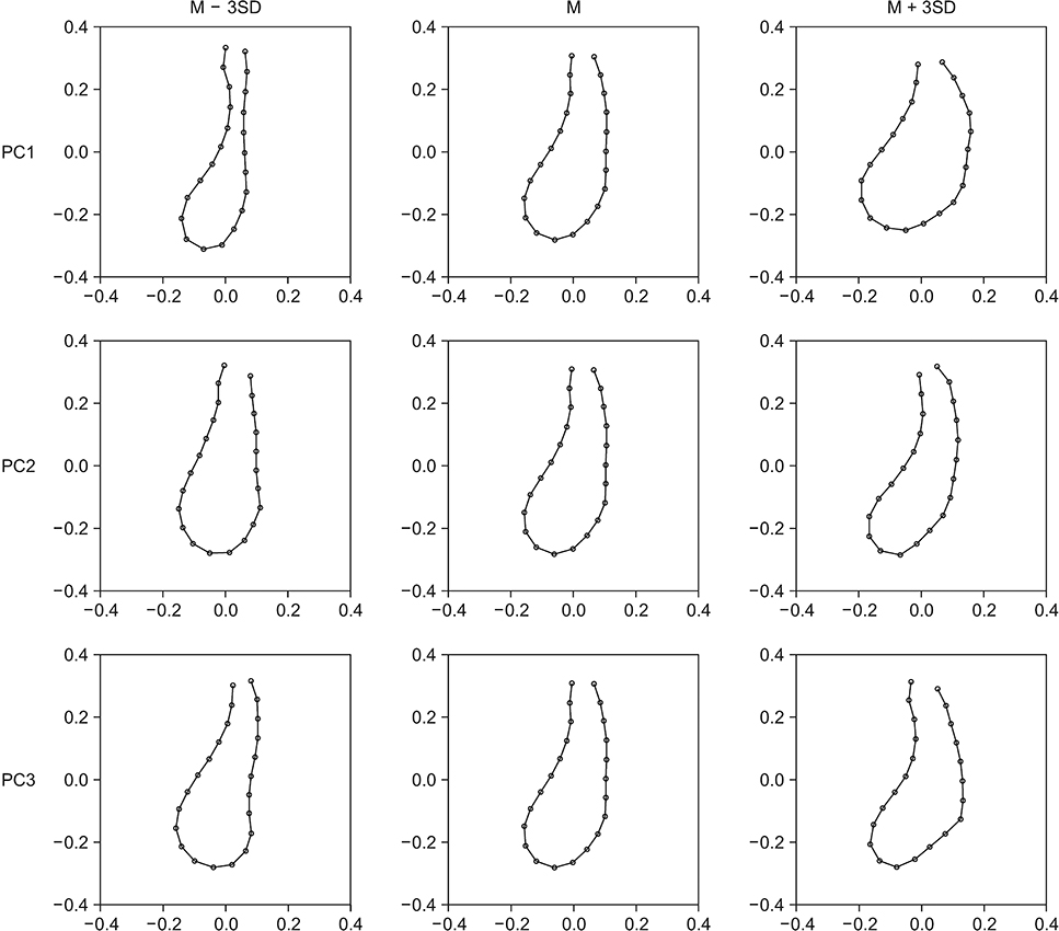
Figure 3 Principal component (PC) analysis of the external cortices of the mandibular symphysis. M, Mean; SD, standard deviation.
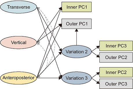
Figure 4 Structural equation model of the skeletal factors and mandibular cortices of the symphysis. The double and dotted arrows show the relationship of the vertical, horizontal, and anteroposterior latent variables. PC, Principal component.
Reference
-
1. Khan MY, Kishore MS, Bukhari SA, Rachala MR, Sashidhar NR. Alveolar and skeletal chin dimensions associated with lower facial height among different divergent patterns. J Clin Diagn Res. 2016; 10:ZC75–ZC80.
Article2. Haskell BS. The human chin and its relationship to mandibular morphology. Angle Orthod. 1979; 49:153–166.3. Kubota M, Nakano H, Sanjo I, Satoh K, Sanjo T, Kamegai T, et al. Maxillofacial morphology and masseter muscle thickness in adults. Eur J Orthod. 1998; 20:535–542.
Article4. Eröz UB, Ceylan I, Aydemir S. An investigation of mandibular morphology in subjects with different vertical facial growth patterns. Aust Orthod J. 2000; 16:16–22.5. Ceylan I, Eröz UB. The effects of overbite on the maxillary and mandibular morphology. Angle Orthod. 2001; 71:110–115.6. Beckmann SH, Kuitert RB, Prahl-Andersen B, Segner D, The RP, Tuinzing DB. Alveolar and skeletal dimensions associated with overbite. Am J Orthod Dentofacial Orthop. 1998; 113:443–452.7. Nojima K, Nakakawaji K, Sakamoto T, Isshiki Y. Relationships between mandibular symphysis morphology and lower incisor inclination in skeletal class III malocclusion requiring orthognathic surgery. Bull Tokyo Dent Coll. 1998; 39:175–181.8. Shimomoto Y, Chung CJ, Iwasaki-Hayashi Y, Muramoto T, Soma K. Effects of occlusal stimuli on alveolar/jaw bone formation. J Dent Res. 2007; 86:47–51.
Article9. Garn SM, Lewis AB, Vicinus JH. The inheritance of symphyseal size during growth. Angle Orthod. 1963; 33:222–231.10. Diedrich P. [Problems and risks in the movement of the mandibular anterior teeth]. Fortschr Kieferorthop. 1995; 56:148–156. German.11. Handelman CS. The anterior alveolus: its importance in limiting orthodontic treatment and its influence on the occurrence of iatrogenic sequelae. Angle Orthod. 1996; 66:95–109. discussion 109-10.12. Aki T, Nanda RS, Currier GF, Nanda SK. Assessment of symphysis morphology as a predictor of the direction of mandibular growth. Am J Orthod Dentofacial Orthop. 1994; 106:60–69.
Article13. Ricketts RM. Cephalometric synthesis. Am J Orthod Dentofacial Orthop. 1960; 46:647–673.
Article14. Sassouni V. A classification of skeletal facial types. Am J Orthod. 1969; 55:109–123.
Article15. Esenlik E, Sabuncuoglu FA. Alveolar and symphysis regions of patients with skeletal class II division 1 anomalies with different vertical growth patterns. Eur J Dent. 2012; 6:123–132.
Article16. Wang MF, Otsuka T, Akimoto S, Sato S. Vertical facial height and its correlation with facial width and depth: three dimensional cone beam computed tomography evaluation based on dry skulls. Int J Stomatol Occlusion Med. 2013; 6:120–129.17. Isaacson JR, Isaacson RJ, Speidel TM, Worms FW. Extreme variation in vertical facial growth and associated variation in skeletal and dental relations. Angle Orthod. 1971; 41:219–229.18. Molina-Berlanga N, Llopis-Perez J, Flores-Mir C, Puigdollers A. Lower incisor dentoalveolar compensation and symphysis dimensions among Class I and III malocclusion patients with different facial vertical skeletal patterns. Angle Orthod. 2013; 83:948–955.
Article19. Al-Khateeb SN, Al Maaitah EF, Abu Alhaija ES, Badran SA. Mandibular symphysis morphology and dimensions in different anteroposterior jaw relationships. Angle Orthod. 2014; 84:304–309.
Article20. Swasty D, Lee J, Huang JC, Maki K, Gansky SA, Hatcher D, et al. Cross-sectional human mandibular morphology as assessed in vivo by cone-beam computed tomography in patients with different vertical facial dimensions. Am J Orthod Dentofacial Orthop. 2011; 139:4 Suppl. e377–e389.
Article
- Full Text Links
-
- Actions
-
Cited
- CITED
-
- Close
- Share
-
- Similar articles
-
- A study on the relationship of the mandibular symphysis and anterior alveolar and skeletal morphology according to the rotational growth pattern of mandible in skeletal Class III malocclusion
- Morphology of mandibular symphysis and positioning of lower incisors in the skeletal Class III malocclusions
- A study of morphoiogy of mandibular symphysis and location of lower incisor under the influence of the craniofacial skeleton in skeletal Class III malocclusion
- A study on correlationship between craniofacial growth pattern and symphysis morphology
- Changes of symphysis morphology after chincup treatment
