In vivo optical coherence tomographic imaging to monitor gingival recovery and the adhesive interface in aesthetic oral rehabilitation: A case report
- Affiliations
-
- 1Department of Prosthodontics and Bucco-Facial Surgery, School of Dentistry, Federal University of Pernambuco, Recife, Brazil. claudio_rec@hotmail.com
- 2Department of Dentistry, UNINASSAU, Caruaru, Brazil.
- 3Department of Biomaterials and Oral Biology, School of Dentistry, University of São Paulo, São Paulo, Brazil.
- 4Department of Physics, Federal University of Pernambuco - UFPE, Recife, Brazil.
- KMID: 2450183
- DOI: http://doi.org/10.5624/isd.2019.49.2.171
Abstract
- The available methods for veneer evaluation are limited to clinical and radiographic examinations, which may not allow the appropriate identification of failure. In this report, we demonstrate the use of optical coherence tomography (OCT) as a noninvasive diagnostic and follow-up method to evaluate gingival recovery and the adhesive interface in aesthetic oral rehabilitation involving periodontal plastic surgery and ceramic laminate veneers. OCT was efficient for evaluating both soft and hard tissues, as well as the quality of the adhesive interface. In conclusion, OCT was found to be a promising approach for the professional evaluation of aesthetic oral rehabilitation, as it was capable of generating images that enabled the analysis of gingival recovery and the adhesive interface.
Keyword
MeSH Terms
Figure
-
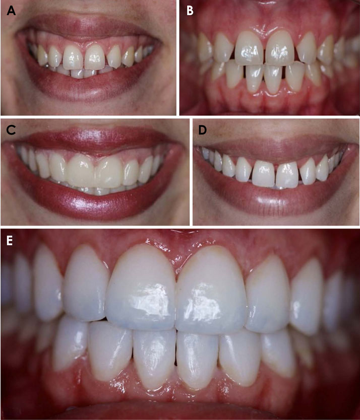
Fig. 1 Clinical photographs of the patient. Extraoral (A) and intraoral (B) photographs show the presence of yellowish teeth, generalized anterior diastema, and a gummy smile. C. Mock-up placed on teeth for the clinical simulation of the treatment outcome. The treatment consisted of periodontal plastic surgery, tooth whitening, and cementation of ceramic veneers. D. View of the patient's smile after periodontal plastic surgery and tooth whitening. E. Immediate clinical view after the cementation of the veneers.
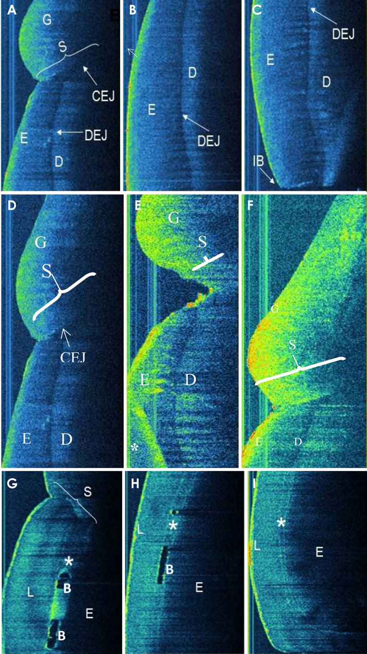
Fig. 2 Sagittal optical coherence tomography images. The cervical (A), middle (B), and incisal (C) thirds of the right maxillary central incisor showing a normal tooth and its surrounding periodontal structures, such as enamel (E), dentin (D), the dentino-enamel junction (DEJ), the cemento-enamel junction (CEJ), gingiva (G), gingival sulcus (S), and incisal border (IB). D. Cervical third of the tooth at 15 days (E) and 60 days (F) after the periodontal plastic surgery demonstrating gingival recovery, given the formation of a new gingival sulcus around the teeth and close contact of the gingiva with the tooth, as shown before surgery. The cervical (G), middle (H), and incisal (I) thirds of the right maxillary central incisor after cementation of the ceramic veneers showing the tooth and its surrounding periodontal structures, as well as the presence of the cementation line (*), bubbles (B), and the laminate veneer (L).
Reference
-
1. Van der Geld P, Oosterveld P, Van Heck G, Kuijpers-Jagtman AM. Smile attractiveness. Self-perception and influence on personality. Angle Orthod. 2007; 77:759–765.2. Morita RK, Hayashida MF, Pupo YM, Berger G, Reggiani RD, Betiol EA. Minimally invasive laminate veneers: clinical aspects in treatment planning and cementation procedures. Case Rep Dent. 2016; 2016:1839793.
Article3. Garcia-Godoy F, Krämer N, Feilzer AJ, Frankenberger R. Long-term degradation of enamel and dentin bonds: 6-year results in vitro vs. in vivo. Dent Mater. 2010; 26:1113–1118.
Article4. Hayashi J, Shimada Y, Tagami J, Sumi Y, Sadr A. Real-time imaging of gap progress during and after composite polymerization. J Dent Res. 2017; 96:992–998.
Article5. Campos F, Valandro LF, Feitosa SA, Kleverlaan CJ, Feilzer AJ, de Jager N, et al. Adhesive cementation promotes higher fatigue resistance to zirconia crowns. Oper Dent. 2017; 42:215–224.
Article6. Huang D, Swanson EA, Lin CP, Schuman JS, Stinson WG, Chang W, et al. Optical coherence tomography. Science. 1991; 254:1178–1181.
Article7. Al-Mujaini A, Wali UK, Azeem S. Optical coherence tomography: clinical applications in medical practice. Oman Med J. 2013; 28:86–91.
Article8. Moraes Pinto Blumetti TC, Cohen MP, Gomes EE, Petaccia de Macedo M, Ferreira de Souza Begnami MD, Tavares Guerreiro Fregnani JH, et al. Optical coherence tomography (OCT) features of nevi and melanomas and their association with intraepidermal or dermal involvement: a pilot study. J Am Acad Dermatol. 2015; 73:315–317.9. John R, Adie SG, Chaney EJ, Marjanovic M, Tangella KV, Boppart SA. Three-dimensional optical coherence tomography for optical biopsy of lymph nodes and assessment of metastatic disease. Ann Surg Oncol. 2013; 20:3685–3693.
Article10. Lopez AL 3rd, Wang S, Larin KV, Overbeek PA, Larina IV. Live four-dimensional optical coherence tomography reveals embryonic cardiac phenotype in mouse mutant. J Biomed Opt. 2015; 20:090501.11. Hsieh YS, Ho YC, Lee SY, Chuang CC, Tsai JC, Lin KF, et al. Dental optical coherence tomography. Sensors (Basel). 2013; 13:8928–8949.
Article12. de Melo LS, de Araujo RE, Freitas AZ, Zezell D, Vieira ND, Girkin J, et al. Evaluation of enamel dental restoration interface by optical coherence tomography. J Biomed Opt. 2005; 10:064027.
Article13. Braz AK, Kyotoku BB, Braz R, Gomes AS. Evaluation of crack propagation in dental composites by optical coherence tomography. Dent Mater. 2009; 25:74–79.
Article14. Alhekeir DF, Al-Sarhan RA, Al Mashaan AF. Porcelain laminate veneers: clinical survey for evaluation of failure. Saudi Dent J. 2014; 26:63–67.
Article15. Machoy M, Seeliger J, Szyszka-Sommerfeld L, Koprowski R, Gedrange T, Woźniak K. The use of optical coherence tomography in dental diagnostics: a state-of-the-art review. J Healthc Eng. 2017; 2017:7560645.
Article16. Xiang X, Sowa MG, Iacopino AM, Maev RG, Hewko MD, Man A, et al. An update on novel non-invasive approaches for periodontal diagnosis. J Periodontol. 2010; 81:186–198.
Article17. Reddy MS. Achieving gingival esthetics. J Am Dent Assoc. 2003; 134:295–304.
Article18. Vanlıoğlu BA, Kulak-Özkan Y. Minimally invasive veneers: current state of the art. Clin Cosmet Investig Dent. 2014; 6:101–107.19. Morimoto S, Albanesi RB, Sesma N, Agra CM, Braga MM. Main clinical outcomes of feldspathic porcelain and glass-ceramic laminate veneers: a systematic review and meta-analysis of survival and complication rates. Int J Prosthodont. 2016; 29:38–49.
Article20. Silva LHD, Lima E, Miranda RB, Favero SS, Lohbauer U, Cesar PF. Dental ceramics: a review of new materials and processing methods. Braz Oral Res. 2017; 31:suppl 1. e58.
Article21. Chadwick RG, McCabe JF, Carrick TE. Rheological properties of veneer trial pastes relevant to clinical success. Br Dent J. 2008; 204:E11.
Article22. Rodrigues RB, Lima E, Roscoe MG, Soares CJ, Cesar PF, Novais VR. Influence of resin cements on color stability of different ceramic systems. Braz Dent J. 2017; 28:191–195.
Article23. Pissaia JF, Correr GM, Gonzaga CC, da Cunha LF. Influence of shade, curing mode, and aging on the color stability of resin cements. Braz J Oral Sci. 2015; 14:272–275.
Article24. de Andrade Borges E, Cassimiro-Silva PF, Fernandes LO, Gomes AS. Study of lumineers' interfaces by means of optical coherence tomography. Proc. SPIE Biophotonics South America. 2015; 9531:953147.25. Hall A, Girkin JM. A review of potential new diagnostic modalities for caries lesions. J Dent Res. 2004; 83:C89–C94.
Article
- Full Text Links
-
- Actions
-
Cited
- CITED
-
- Close
- Share
-
- Similar articles
-
- Usefulness of Optical Coherence Tomography in the Preoperative Assessment of Nail Deformities
- Early caries detection using optical coherence tomography: a review of the literature
- Applications of Optical Imaging System in Dentistry
- Optical Imaging and Its Clinical Application in Otorhinolaryngology
- Photoacoustic imaging platforms for multimodal imaging
