Pneumatosis Intestinalis Presenting as Small Bowel Obstruction without Bowel Ischemia after Mechanical Ventilation
- Affiliations
-
- 1Department of Anesthesiology and Pain Medicine, Chosun University Hospital, Gwangju, Korea.
- 2Department of Surgery, Chosun University Hospital, Gwangju, Korea. ysyoo@chosun.ac.kr
- KMID: 2449384
- DOI: http://doi.org/10.4266/acc.2016.00311
Abstract
- Pneumatosis intestinalis (PI) is a rare condition of the presence of gas within the bowel walls. PI is associated with numerous underlying diseases, ranging from life-threatening to innocuous conditions. PI is believed to be secondary to coexisting disorders in approximately 85% of all cases. This paper reviews the case of a patient who was diagnosed 7 years prior with pneumoperitoneum from unknown causes without any symptoms. The patient was admitted to the intensive care unit for the management of aspiration pneumonia and developed extensive PI after mechanical ventilation, presenting as small bowel obstruction with mesenteric torsion. Although the exact mechanism and etiology of PI are unclear, this case provides an update on the imaging features of and the clinical conditions associated with PI, as well as the management of this condition.
MeSH Terms
Figure
-
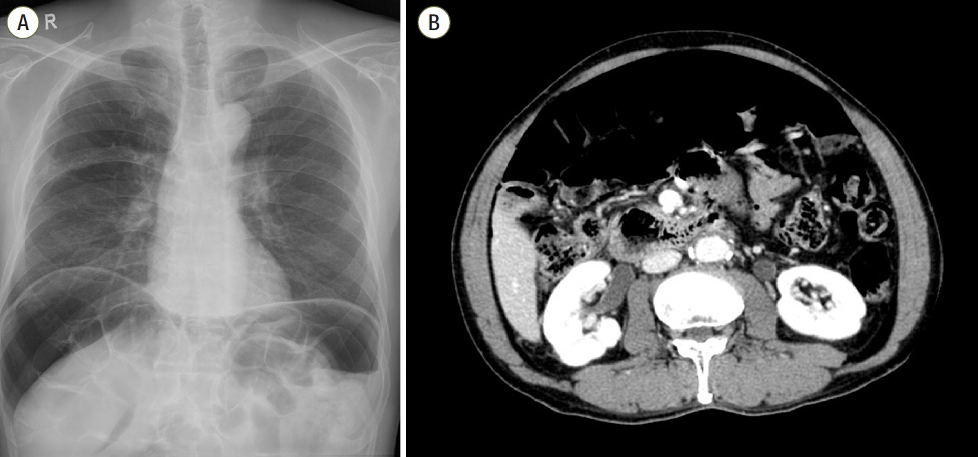
Figure 1. (A) Massive free air at both subdiaphragmatic areas. Mildly emphysematous lung on the right. (B) Large amount of free air in the abdominal cavity on computed tomography. Both Figures taken 7 years prior.
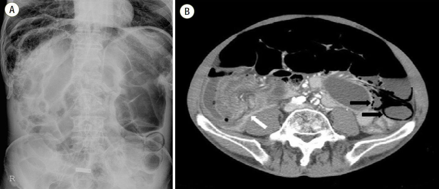
Figure 2. (A) Septated pneumoperitoneum at the right subphrenic area. Dilatation and pneumatosis intestinalis in the small bowel. (B) Segmental edematous small bowel wall thickening with air bubbles (black arrows), and mesenteric torsion showing a whirling sign (white arrow).
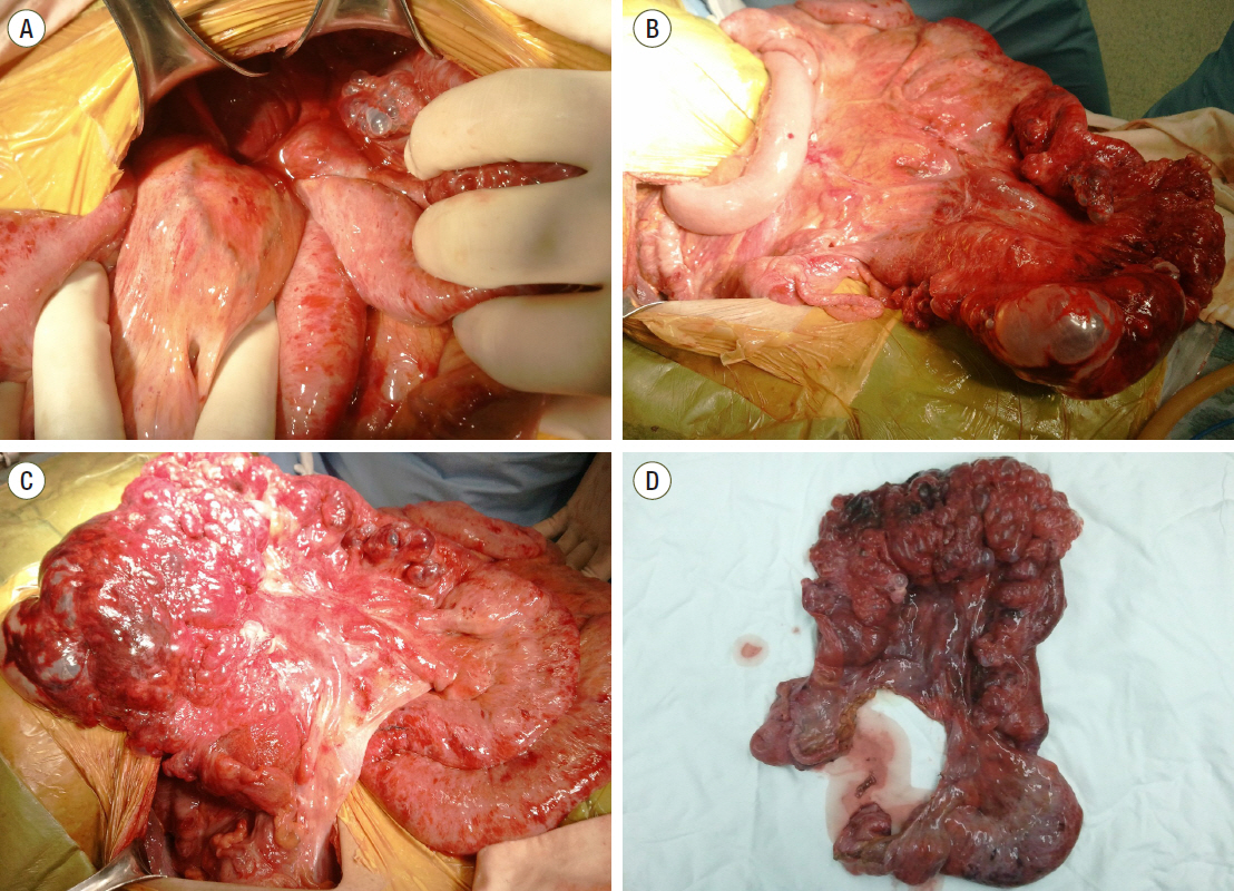
Figure 3. (A) Twisted small bowel loops in the right lower quadrant. (B) Reduction of stacked small bowel showing massive pneumatosis intestinalis. (C) The other side of the small bowel mesentery showing large bubbles on the small bowel wall. (D) Resected specimen showing extensive pneumatosis intestinalis.
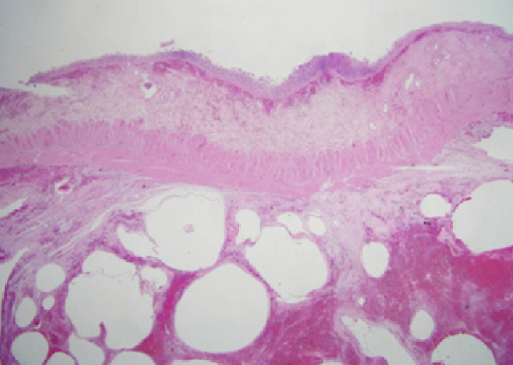
Figure 4. H&E staining of small bowel (×5). Numerous air pockets were observed in the subserosal layer.
Reference
-
1. Morris MS, Gee AC, Cho SD, Limbaugh K, Underwood S, Ham B, et al. Management and outcome of pneumatosis intestinalis. Am J Surg. 2008; 195:679–82.
Article2. Koss LG. Abdominal gas cysts (pneumatosis cystoides intetsinorum hominis); an analysis with a report of a case and a critical review of the literature. AMA Arch Pathol. 1952; 53:523–49.3. St Peter SD, Abbas MA, Kelly KA. The spectrum of pneumatosis intestinalis. Arch Surg. 2003; 138:68–75.
Article4. Heng Y, Schuffler MD, Haggitt RC, Rohrmann CA. Pneumatosis intestinalis: a review. Am J Gastroenterol. 1995; 90:1747–58.5. Braumann C, Menenakos C, Jacobi CA. Pneumatosis intestinalis: a pitfall for surgeons? Scand J Surg. 2005; 94:47–50.6. Jamart J. Pneumatosis cystoides intestinalis: a statistical study of 919 cases. Acta Hepatogastroenterol (Stuttg). 1979; 26:419–22.7. Greenstein AJ, Nguyen SQ, Berlin A, Corona J, Lee J, Wong E, et al. Pneumatosis intestinalis in adults: management, surgical indications, and risk factors for mortality. J Gastrointest Surg. 2007; 11:1268–74.
Article8. Kim HL, Lee HK, Park SJ, Yi BH, Ko BM, Hong HS, et al. Pneumatosis intestinalis: CT findings and clinical features. J Korean Radiol Soc. 2008; 58:149–54.
Article9. Donovan S, Cernigliaro J, Dawson N. Pneumatosis intestinalis: a case report and approach to management. Case Rep Med. 2011; 2011:571387.
Article10. Brant WE. Fundamentals of diagnostic radiology. 4th ed. Philadelphia: Wolters Kluwer Health;2012.11. Webb WR. Fundamentals of body CT. 2nd ed. Philadelphia: Elsevier Health Sciences;2014.
- Full Text Links
-
- Actions
-
Cited
- CITED
-
- Close
- Share
-
- Similar articles
-
- A Rare Case of Hypermobile Mesentery With Segmental Small Bowel Pneumatosis Cystoides Intestinalis
- A Case of Necrotizing Colitis Presenting with Hepatic Portal Venous Gas and Pneumatosis Intestinalis
- A Case of Pneumatosis Cystoides Intestinalis in a Patient with Chronic Diarrhea and Abdominal Pain
- A Case of Pneumatosis Intestinalis in Peritoneal Dialysis Peritonitis
- Pneumatosis Cystoides Intestinalis and Partial Abdominal Obstruction in a Patient with Polymyositis
