A Removed Chestnut Thorn after Corneal Incision
- Affiliations
-
- 1Department of Ophthalmology, Pusan National University Yangsan Hospital, Pusan National University School of Medicine, Yangsan, Korea. jiel@hanmail.net
- KMID: 2445146
- DOI: http://doi.org/10.3341/jkos.2019.60.5.496
Abstract
- PURPOSE
To report the surgical technique to remove a chestnut thorn through a corneal incision.
CASE SUMMARY
A 54-year-old female visited our clinic complaining of a sudden foreign body sensation and conjunctival injection in her left eye after picking chestnuts 4 days prior to her visit. Visual acuity of both eyes was 1.0 and the intraocular pressures were within normal limits. Slit lamp examination revealed that a chestnut thorn had deeply penetrated the left corneal stroma and a small number of inflammatory cells were observed in the anterior chamber. There was no corneal defect stained with fluorescein and the Seidel test was negative. A corneal foreign body comprised of a chestnut thorn and its remnants was diagnosed and emergency surgery was performed. A partial corneal incision was made along the foreign body and the exposed foreign body was easily and completely removed. The patient was treated with topical antibiotics after surgery and no complication was observed during a follow-up period of 3 months.
CONCLUSIONS
In the case of a corneal foreign body comprised of a chestnut thorn, the foreign body with its remnants were easily removed by performing a partial corneal incision.
Keyword
MeSH Terms
Figure
-
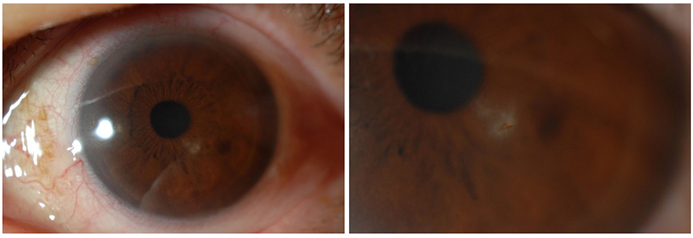
Figure 1 Slit-lamp ophthalmoscopic findings at initial visit. Chestnut thorn penetrated the cornea.
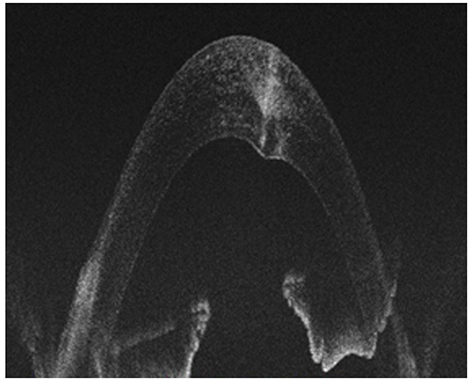
Figure 2 Anterior segment optical coherence tomography at initial visit. The chestnut thorn penetrated the cornea deep into the Descemet's membrane.
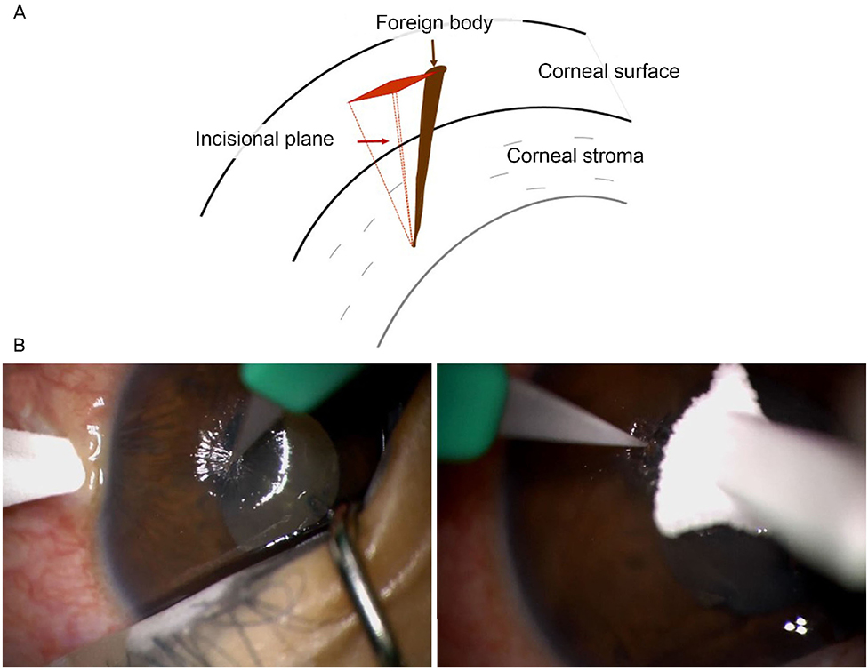
Figure 3 Surgical procedure to remove chestnut thorn through the corneal incision. (A) Schematic image of removing foreign body from the cornea. The foreign body was deeply embedded into the corneal stroma, and a partial corneal incision was made along the foreign body. (B) After corneal incision, the deepest part of the foreign body was reversely pushed to the outside of the cornea with the knife.
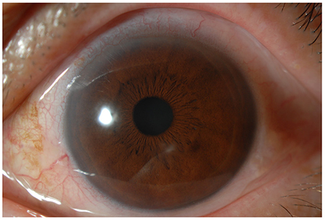
Figure 4 Slit-lamp findings of cornea at 3 months after the surgery. Mild stromal opacity was remained at the previous foreign body site.
Reference
-
1. Oh SU, Kim TY. Clinical evaluation of chestnut bur injuries to the eye. J Korean Ophthalmol Soc. 2000; 41:2174–2179.2. Duan H, Yan S. Clinical efficacy of surgical removal of deep corneal plant foreign bodies. Eye Sci. 2013; 28:30–33.3. Bradford GE, Burnstine RA. A technique for removing thorns from the cornea. J Pediatr Ophthalmol Strabismus. 1992; 29:319.
Article4. Jung JH, Kim O, Kim HB, et al. A statistical observation of the corneal foreign bodies. J Korean Ophthalmol Soc. 1972; 13:153–156.5. Macedo Filho ET, Lago A, Duarte K, et al. Superficial corneal foreign body: laboratory and epidemiologic aspects. Arq Bras Oftalmol. 2005; 68:821–823.
Article6. Kim MJ, Lee U, Hwang MS, et al. Blooming, fructification and nut characteristics of chestnut cultivars in Korea. J Korean For Soc. 2003; 92:321–332.7. Chen WL, Tseng CH, Wang IJ, Hu FR. Removal of semitranslucent cactus spines embedded in deep cornea with the aid of a fiberoptic illuminator. Am J Ophthalmol. 2002; 134:769–771.
Article
- Full Text Links
-
- Actions
-
Cited
- CITED
-
- Close
- Share
-
- Similar articles
-
- Changes in Corneal Sensitivity after Cataract Surgery
- Rupture of the extensor digitorum communis tendon in extensor zone V due to plant thorn injuries: a report of two cases
- The Corneal Topographic Changes in Sutureless Cataract Surgery
- Mid Limbal Incision vs Scleral Pocket Incision in Cataract Surgery
- The Location of Scleral Incision for Decrease of corneal Astigmatism in Sutureless Cataract Surgery
