Comparison of Blue and Green Confocal Scanning Laser Ophthalmoscope Imaging to Detect Retinal Nerve Fiber Layer Defects
- Affiliations
-
- 1Department of Ophthalmology, Hanyang University College of Medicine, Seoul, Korea. brlee@hanyang.ac.kr
- KMID: 2442617
- DOI: http://doi.org/10.3341/kjo.2018.0075
Abstract
- PURPOSE
We detected retinal nerve fiber layer (RNFL) defects using a confocal scanning laser ophthalmoscopy (CSLO) with both blue and green laser sources and evaluated image quality based on laser wavelength.
METHODS
This was a retrospective observational case study. Blue and green CSLO images of 181 eyes with suspected glaucoma were evaluated and compared. Three independent observers identified the presence of RNFL defects and determined which CSLO imaging source provided superior visibility of the defect. After assessing the defect imaging by laser source, demographics and image quality indices of optical coherence tomography between blue better and green better groups were analyzed.
RESULTS
Both blue and green CSLO showed high discriminating ability for RNFL defects. The discriminating ability of blue CSLO was significantly greater than that of green CSLO (p = 0.004). Among eyes with a detectable RNFL defect, 61.8% were better visualized with the blue laser compared to the green laser. Compared with the blue better group, the green better group was significantly older (p = 0.009), had a greater proportion of females (p = 0.005), had poorer best-corrected visual acuity (p = 0.001), more severe cataracts (p = 0.001), lower signal strength (p = 0.003), and poor image quality indices (p = 0.001).
CONCLUSIONS
Both blue and green CSLO imaging was useful for detecting RNFL defects, but blue CSLO was superior to green CSLO in quality of RNFL defect imaging in most patients with clear media.
MeSH Terms
Figure
-
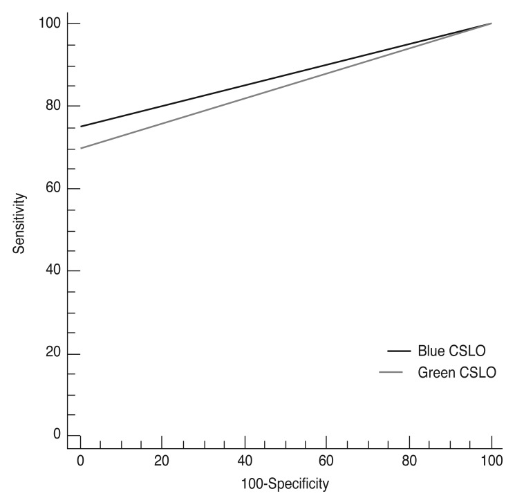
Fig. 1 Comparison of receiver operating characteristic curves created using blue confocal scanning laser ophthalmoscope (CSLO) imaging and green CSLO imaging.
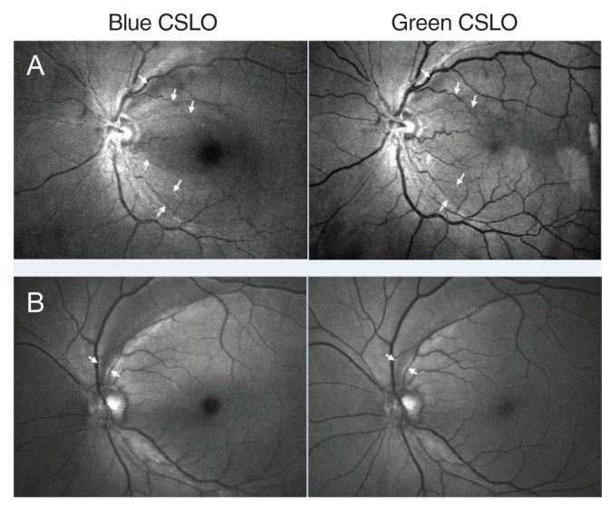
Fig. 2 Representative confocal scanning laser ophthalmoscope (CSLO) imaging of the blue better group. (A) Case 1: 82-year-old male. Multiple retinal nerve fiber layer (RNFL) defects (white arrow) were visible with both blue and green CSLO imaging. Visualization of RNFL defects was better with blue compared to green CSLO imaging. (B) Case 2: 63-year-old female. A superior temporal RNFL defect (white arrow) was visible with both blue and green CSLO imaging. Visualization of the RNFL defect was better with blue compared to green CSLO imaging.

Fig. 3 Representative confocal scanning laser ophthalmoscope (CSLO) imaging of the green better group. (A) Case 3: 73-year-old female. An inferior temporal retinal nerve fiber layer (RNFL) defect (white arrow) was visible with both blue and green CSLO imaging. This patient had corneal opacity. Visualization of the RNFL defect was better with green versus blue CSLO imaging. (B) Case 4: 76-year-old female. An inferior RNFL defect (white arrow) was visible with both blue and green CSLO imaging. The patient had severe cataracts according to slit lamp examination. Visualization of the RNFL defect was better with green versus blue CSLO imaging. In particular, the boundary of the RNFL defect and its relationship with adjacent retinal vasculature were more clearly visible with green CSLO imaging.
Reference
-
1. Hoyt WF, Frisen L, Newman NM. Fundoscopy of nerve fiber layer defects in glaucoma. Invest Ophthalmol. 1973; 12:814–829. PMID: 4752920.2. Quigley HA, Katz J, Derick RJ, et al. An evaluation of optic disc and nerve fiber layer examinations in monitoring progression of early glaucoma damage. Ophthalmology. 1992; 99:19–28. PMID: 1741133.
Article4. Sommer A, Katz J, Quigley HA, et al. Clinically detectable nerve fiber atrophy precedes the onset of glaucomatous field loss. Arch Ophthalmol. 1991; 109:77–83. PMID: 1987954.
Article5. Sommer A, Miller NR, Pollack I, et al. The nerve fiber layer in the diagnosis of glaucoma. Arch Ophthalmol. 1977; 95:2149–2156. PMID: 588106.
Article6. Jonas JB, Schiro D. Localised wedge shaped defects of the retinal nerve fibre layer in glaucoma. Br J Ophthalmol. 1994; 78:285–290. PMID: 8199115.
Article7. Tuulonen A, Airaksinen PJ, Montagna A, Nieminen H. Screening for glaucoma with a non-mydriatic fundus camera. Acta Ophthalmol (Copenh). 1990; 68:445–449. PMID: 2220362.
Article8. Townsend KA, Wollstein G, Schuman JS. Imaging of the retinal nerve fibre layer for glaucoma. Br J Ophthalmol. 2009; 93:139–143. PMID: 19028735.
Article9. DeLong ER, DeLong DM, Clarke-Pearson DL. Comparing the areas under two or more correlated receiver operating characteristic curves: a nonparametric approach. Biometrics. 1988; 44:837–845. PMID: 3203132.
Article10. Behrendt T, Wilson LA. Spectral reflectance photography of the retina. Am J Ophthalmol. 1965; 59:1079–1088. PMID: 14292719.11. Miller NR, George TW. Monochromatic (red-free) photography and ophthalmoscopy of the peripapillary retinal nerve fiber layer. Invest Ophthalmol Vis Sci. 1978; 17:1121–1124. PMID: 700962.12. Peli E, Hedges TR 3rd, McInnes T, et al. Nerve fiber layer photography: a comparative study. Acta Ophthalmol (Copenh). 1987; 65:71–80. PMID: 3577709.13. Frisen L. Photography of the retinal nerve fibre layer: an optimized procedure. Br J Ophthalmol. 1980; 64:641–650. PMID: 7426584.14. Airaksinen PJ, Nieminen H, Mustonen E. Retinal nerve fibre layer photography with a wide angle fundus camera. Acta Ophthalmol (Copenh). 1982; 60:362–368. PMID: 7136547.
Article15. Sommer A, D'Anna SA, Kues HA, George T. High-resolution photography of the retinal nerve fiber layer. Am J Ophthalmol. 1983; 96:535–539. PMID: 6624835.
Article16. Airaksinen PJ, Drance SM, Douglas GR, et al. Diffuse and localized nerve fiber loss in glaucoma. Am J Ophthalmol. 1984; 98:566–571. PMID: 6496612.
Article17. Airaksinen PJ, Nieminen H. Retinal nerve fiber layer photography in glaucoma. Ophthalmology. 1985; 92:877–879. PMID: 4022571.
Article18. Hitchings RA, Poinoosawmy D, Poplar N, Sheth GP. Retinal nerve fibre layer photography in glaucomatous patients. Eye (Lond). 1987; 1:621–625. PMID: 3446544.
Article19. Han ES, Park KH. Using red-free monochromatic conversions of nonmydriatic digital fundus images. Am J Ophthalmol. 2007; 143:371–372. PMID: 17258543.
Article20. Hong S, Ahn H, Ha SJ, et al. Early glaucoma detection using the Humphrey Matrix Perimeter, GDx VCC, Stratus OCT, and retinal nerve fiber layer photography. Ophthalmology. 2007; 114:210–215. PMID: 17270671.
Article21. Hong S, Moon JW, Ha SJ, et al. Evaluation of a new scoring system for retinal nerve fiber layer photography using HRA1 in 964 eyes. Korean J Ophthalmol. 2007; 21:216–221. PMID: 18063886.
Article22. Ye C, To E, Weinreb RN, et al. Comparison of retinal nerve fiber layer imaging by spectral domain optical coherence tomography and scanning laser ophthalmoscopy. Ophthalmology. 2011; 118:2196–2202. PMID: 21762989.
Article23. Kawaguchi I, Higashide T, Ohkubo S, et al. In vivo imaging and quantitative evaluation of the rat retinal nerve fiber layer using scanning laser ophthalmoscopy. Invest Ophthalmol Vis Sci. 2006; 47:2911–2916. PMID: 16799033.
Article24. Sommer A, Kues HA, D'Anna SA, et al. Cross-polarization photography of the nerve fiber layer. Arch Ophthalmol. 1984; 102:864–869. PMID: 6732566.
Article25. Rohrschneider K, Kruse FE, Burk RO, Volcker HE. Possibilities for imaging the retinal nerve fiber layer with the scanning laser ophthalmoscope. Ophthalmologe. 1995; 92:515–520. PMID: 7549339.26. Beckman C, Bond-Taylor L, Lindblom B, Sjostrand J. Confocal fundus imaging with a scanning laser ophthalmoscope in eyes with cataract. Br J Ophthalmol. 1995; 79:900–904. PMID: 7488577.
Article27. Webb RH, Hughes GW. Scanning laser ophthalmoscope. IEEE Trans Biomed Eng. 1981; 28:488–492. PMID: 7275128.
Article28. Webb RH, Hughes GW, Delori FC. Confocal scanning laser ophthalmoscope. Appl Opt. 1987; 26:1492–1499. PMID: 20454349.
Article
- Full Text Links
-
- Actions
-
Cited
- CITED
-
- Close
- Share
-
- Similar articles
-
- Biometry of Retinal Nerve Fiber Layer Thickness by NFA
- Reproducibility of Retinal Nerve Fiber Layer Thickness Evaluation by Nerve Fiber Analyzer
- Topographic Measurements of the Optic Nerve Head with Confocal Scanning Laser Tomography in Normal Subjects and Patients with Glaucoma
- The Relationship between the Duration of IOP Elevation during LASIK and Nerve Fiber Layer Thickness Measured by GDx(R)
- The Parameters of the Retinal Nerve Fiber Layer Measured with Confocal Scanning Laser Ophthalmoscope & Nerve Fiber Analyzer
