Concha bullosa, nasal septal deviation, and their impacts on maxillary sinus volume among Emirati people: A cone-beam computed tomography study
- Affiliations
-
- 1Department of Oral and Craniofacial Health Sciences, College of Dental Medicine, University of Sharjah, Sharjah, United Arab Emirates. nhabdulla@sharjah.ac.ae
- 2Department of Dental Surgical Sciences, College of Dentistry, Gulf Medical University, Ajman, United Arab Emirates.
- 3Department of Clinical Research-Clinical Affairs Directorate, Primary Health Care Corporation (PHCC), Doha, Qatar.
- KMID: 2442108
- DOI: http://doi.org/10.5624/isd.2019.49.1.45
Abstract
- PURPOSE
To determine the prevalence of concha bullosa (CB) and nasal septal deviation (NSD) and their impact on maxillary sinus volume (MSV).
MATERIALS AND METHODS
Cone-beam computed tomographic (CBCT) images of 106 Emirati people were used in this study. The direction and angle of septal deviation were calculated. The presence of CB, which could be unilateral, contralateral, or bilateral in relation to the direction of NSD, was also recorded. MSV was measured using reconstructed Digital Imaging and Communication in Medicine images on Dolphin 3D imaging software version 11.8 premium (Dolphin Imaging, Chatsworth, CA, USA). P values<0.05 were considered to indicate statistical significance.
RESULTS
CB was detected in 37.7% of the sample; 20.7% of the sample showed single unilateral CB and 16.6% had single bilateral CB. NSD was seen in 74.5% of the sample. In the participants with CB, 45.5% showed mild deviation, 34.4% showed moderate deviation, and only 12.5% showed severe septal deviation. CB, but not NSD, was associated with significantly higher MSV on the affected side (P=0.001).
CONCLUSION
Although NSD was observed in more than two-thirds of the sample and CB was present in more than one-third of the sample, only CB had a significant impact on MSV.
Figure
-

Fig. 1 Measurement of the angle of septal deviation (the angle between the crista galli and the most prominent point of deviation).
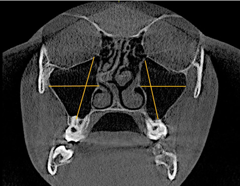
Fig. 2 Measurement of maxillary sinus height (from the lowest point of the sinus floor to the highest point of the sinus roof) and width (from the longest perpendicular distance from the medial wall of the sinus to the outermost point of the lateral wall of the maxillary sinus).

Fig. 3 Measurement of the anteroposterior dimension of the maxillary sinus from the most anterior point to the most posterior point.

Fig. 4 Reconstructed 3-dimensional image of the maxillary sinuses for volume measurements.

Fig. 5 Bilateral concha bullosa from a coronal slice of cone-beam computed tomography.
Cited by 1 articles
-
Prevalence of the anatomical variations of concha bullosa and its relation with sinusitis among Saudi population: a computed tomography scan study
Wael Amin Nasr El-Din, Gisma Ahmed Madani, Islam Omar Abdel Fattah, Esmat Mahmoud, Asmaa S. Essawy
Anat Cell Biol. 2021;54(2):193-201. doi: 10.5115/acb.20.247.
Reference
-
1. Uthman AT, Al-Rawi NH, AL-Naaimi AS, Al-Timimi JF. Evaluation of maxillary sinus dimensions in gender determination using helical CT scanning. J Forensic Sci. 2011; 56:403–408.
Article2. Uthman AT, AL-Rawi NH, Al-Naaimi AS, Tawfeeq AS, Suhail EH. Evaluation of frontal sinus and skull measurements using spiral CT scanning: an aid in unknown person identification. Forensic Sci Int. 2010; 197:124.e1–124.e7.
Article3. Bolger WE, Butzin CA, Parsons DS. Paranasal sinus bony anatomic variations and mucosal abnormalities: CT analysis for endoscopic sinus surgery. Laryngoscope. 1991; 101:56–64.4. Tonai A, Baba S. Anatomic variations of the bone in sinonasal CT. Acta Otolaryngol Suppl. 1996; 525:9–13.5. Aktas D, Kalcioglu MT, Kutlu R, Ozturan O, Oncel S. The relationship between the concha bullosa, nasal septal deviation and sinusitis. Rhinology. 2003; 41:103–106.6. Earwaker J. Anatomic variants in sinonasal CT. Radiographics. 1993; 13:381–415.
Article7. Zinreich SJ, Mattox DE, Kennedy DW, Chisholm HL, Diffley DM, Rosenbaum AE. Concha bullosa: CT evaluation. J Comput Assist Tomogr. 1988; 12:778–784.8. Yiğit O, Acioğlu E, Cakir ZA, Sişman AS, Barut AY. Concha bullosa and septal deviation. Eur Arch Otorhinolaryngol. 2010; 267:1397–1401.
Article9. Hatipoğlu H, Çetin M, Yüksel E. Concha bullosa types: their relationship with sinusitis, ostiomeatal and frontal recess disease. Diagn Interv Radiol. 2005; 11:145–149.10. Alharethy S, Aldrees T, Aljrid R, Alanazi A, Algaryan SK, Jang YJ. Common nasal deformities among rhinoplasty patients in a university hospital in Saudi Arabia. Ann Saudi Med. 2017; 37:2017–2211.
Article11. Wang RG, Jiang SC, Gu R. The cartilaginous nasal capsule and embryonic development of human paranasal sinuses. J Otolaryngol. 1994; 23:239–243.12. Serifoglu I, OZ İİ, Damar M, Buyukuysal M, Tosun A, Tokgöz O. Relationship between the degree and direction of nasal septum deviation and nasal bone morphology. Head Face Med. 2017; 13:3.
Article13. Rak KM, Newell JD 2nd, Yakes WF, Damiano MA, Luethke JM. Paranasal sinuses on MR images of the brain: significance of mucosal thickening. AJR Am J Roentgenol. 1991; 156:381–384.
Article14. Al-Rawi NH, Uthman AT, Sodeify SM. Spatial analysis of mandibular condyles in patients with temporomandibular disorders and normal controls using cone beam computed tomography. Eur J Dent. 2017; 11:99–105.
Article15. Kantarci M, Karasen RM, Alper F, Onbas O, Okur A, Karaman A. Remarkable anatomic variations in paranasal sinus region and their clinical importance. Eur J Radiol. 2004; 50:296–302.
Article16. Shin HS. Clinical significance of unilateral sinusitis. J Korean Med Sci. 1986; 1:69–74.
Article17. Collet S, Bertrand B, Cornu S, Eloy P, Rombaux P. Is septal deviation a risk factor for chronic sinusitis? Review of literature. Acta Otorhinolaryngol Belg. 2001; 55:299–304.18. Kapusuz Gencer Z, Ozkırış M, Okur A, Karaçavuş S, Saydam L. The effect of nasal septal deviation on maxillary sinus volumes and development of maxillary sinusitis. Eur Arch Otorhinolaryngol. 2013; 270:3069–3073.
Article19. Lee DH, Shin JH, Lee DC. Three-dimensional morphometric analysis of paranasal sinuses and mastoid air cell system using computed tomography in pediatric population. Int J Pediatr Otorhinolaryngol. 2012; 76:1642–1646.
Article20. Moorthy PN, Kolloju S, Madhira S, Jowkar AB. Clinical study on deviated nasal septum and its associated pathology. Int J Otolaryngol Head Neck Surg. 2014; 3:75–81.
Article21. Erkan SO, Erkan ZA, Tuhanioğlu B, Haytoğlu S, Güney Z. The relationship between septal deviation and concha bullosa. Kulak Burun Bogaz Ihtis Derg. 2017; 27:74–78.
Article22. Sazgar AA, Massah J, Sadeghi M, Bagheri A, Rasool E. The incidence of concha bullosa and the correlation with nasal septal deviation. B-ENT. 2008; 4:87–91.23. Stallman JS, Lobo JN, Som PM. The incidence of concha bullosa and its relationship to nasal septal deviation and paranasal sinus disease. AJNR Am J Neuroradiol. 2004; 25:1613–1618.24. Calhoun KH, Waggenspack GA, Simpson CB, Hokanson JA, Bailey BJ. CT evaluation of the paranasal sinuses in symptomatic and asymptomatic populations. Otolaryngol Head Neck Surg. 1991; 104:480–483.
Article25. Danese M, Duvoisin B, Agrifoglio A, Cherpillod J, Krayenbuhl M. Influence of naso-sinusal anatomic variants on recurrent, persistent or chronic sinusitis. X-ray computed tomographic evaluation in 112 patients. J Radiol. 1997; 78:651–657.26. Arslan H, Aydinlioglu A, Bozkurt M, Egeli E. Anatomic variations of the paranasal sinuses: CT examination for endoscopic sinus surgery. Auris Nasus Larynx. 1999; 26:39–48.
Article27. Miranda CM, Maranhão CP, Arraes FM, Padilha IG, Farias LP, Jatobá MS, et al. Anatomic variations of paranasal sinuses at multi-slice computed tomography: what to look for. Radiol Bras. 2011; 44:256–262.28. Uygur K, Tüz M, Doğru H. The correlation between septal deviation and concha bullosa. Otolaryngol Head Neck Surg. 2003; 129:33–36.
Article29. Subramanian S, Lekhraj Rampal GR, Wong EF, Mastura S, Razi A. Concha bullosa in chronic sinusitis. Med J Malaysia. 2005; 60:535–539.30. Lee JS, Ko IJ, Kang HD, Lee HS. Massive concha bullosa with secondary maxillary sinusitis. Clin Exp Otorhinolaryngol. 2008; 1:221–223.
Article31. Smith KD, Edwards PC, Saini TS, Norton NS. The prevalence of concha bullosa and nasal septal deviation and their relationship to maxillary sinusitis by volumetric tomography. Int J Dent. 2010; 2010:pii: 404982.
Article32. Göçmen G, Borahan MO, Aktop S, Dumlu A, Pekiner FN, Göker K. Effect of septal deviation, concha bullosa and Haller's cell on maxillary sinus's inferior pneumatization; a retrospective study. Open Dent J. 2015; 9:282–286.
Article33. Demir UL, Akca ME, Ozpar R, Albayrak C, Hakyemez B. Anatomical correlation between existence of concha bullosa and maxillary sinus volume. Surg Radiol Anat. 2015; 37:1093–1098.
Article34. Kucybala I, Janik KA, Ciuk S, Storman D, Urbanik A. Nasal septal deviation and concha bullosa - do they have an impact on maxillary sinus volumes and prevalence of maxillary sinusitis? Pol J Radiol. 2017; 82:126–133.
Article35. Bahemmat N, Hadian H. The frequency of nasal septal deviation and concha bullosa and their relationship with maxillary sinusitis based on CBCT finding. Int J Med Res Health Sci. 2016; 5:152–156.
- Full Text Links
-
- Actions
-
Cited
- CITED
-
- Close
- Share
-
- Similar articles
-
- Does Nasal Septal Deviation and Concha Bullosa Have Effect on Maxillary Sinus Volume and Maxillary Sinusitis?: A Retrospective Study
- Massive Concha Bullosa with Secondary Maxillary Sinusitis
- Concha Bullosa: Incidence and Relationship with Chronic Sinusitis on OMU CT
- The Effect of Radiation Therapy on Paranasal Sinus Opacification in Nasopharyngeal Cancer Patients
- A Case of Giant Concha Bullosa Causing Complete Unilateral Obstruction of Nasal Cavity
