Validation of a low-cost portable 3-dimensional face scanner
- Affiliations
-
- 1Paediatric Dentistry Department, Dental Centre, Ground Floor South Wing, St Thomas' Hospital, Westminster Bridge Road, London, UK. catherineliu.dr@gmail.com
- 2Academic Centre of Reconstructive Science, King's College London Dental Institute, Floor 20, Tower Wing, Guy's Hospital, Great Maze Pond, London, UK.
- KMID: 2442107
- DOI: http://doi.org/10.5624/isd.2019.49.1.35
Abstract
- PURPOSE
The goal of this study was to assess the accuracy and reliability of a low-cost portable scanner (Scanify) for imaging facial casts compared to a previously validated portable digital stereophotogrammetry device (Vectra H1). This in vitro study was performed using 2 facial casts obtained by recording impressions of the authors, at King's College London Academic Centre of Reconstructive Science.
MATERIALS AND METHODS
The casts were marked with anthropometric landmarks, then digitised using Scanify and Vectra H1. Computed tomography (CT) scans of the same casts were performed to verify the validation of Vectra H1. The 3-dimensional (3D) images acquired with each device were compared using linear measurements and 3D surface analysis software.
RESULTS
Overall, 91% of the linear Scanify measurements were within 1 mm of the corresponding reference values. The mean overall surface difference between the Scanify and Vectra images was <0.3 mm. Significant differences were detected in depth measurements. Merging multiple Scanify images produced significantly greater registration error.
CONCLUSION
Scanify is a very low-cost device that could have clinical applications for facial imaging if imaging errors could be corrected by a future software update or hardware revision.
Figure
-

Fig. 1 The experimental device, Scanify (Fuel 3D Technologies, Chinnor, UK).
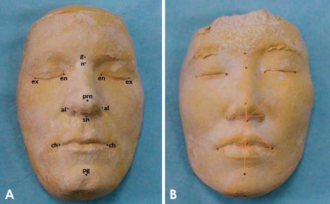
Fig. 2 A. Facial cast (sample 1) illustrating the 13 anthropometric landmarks placed on each cast. B. Facial cast (sample 2) illustrating the 11 inter-landmark measurements that were performed.

Fig. 3 Frontal views of computed tomography scans of both facial casts.
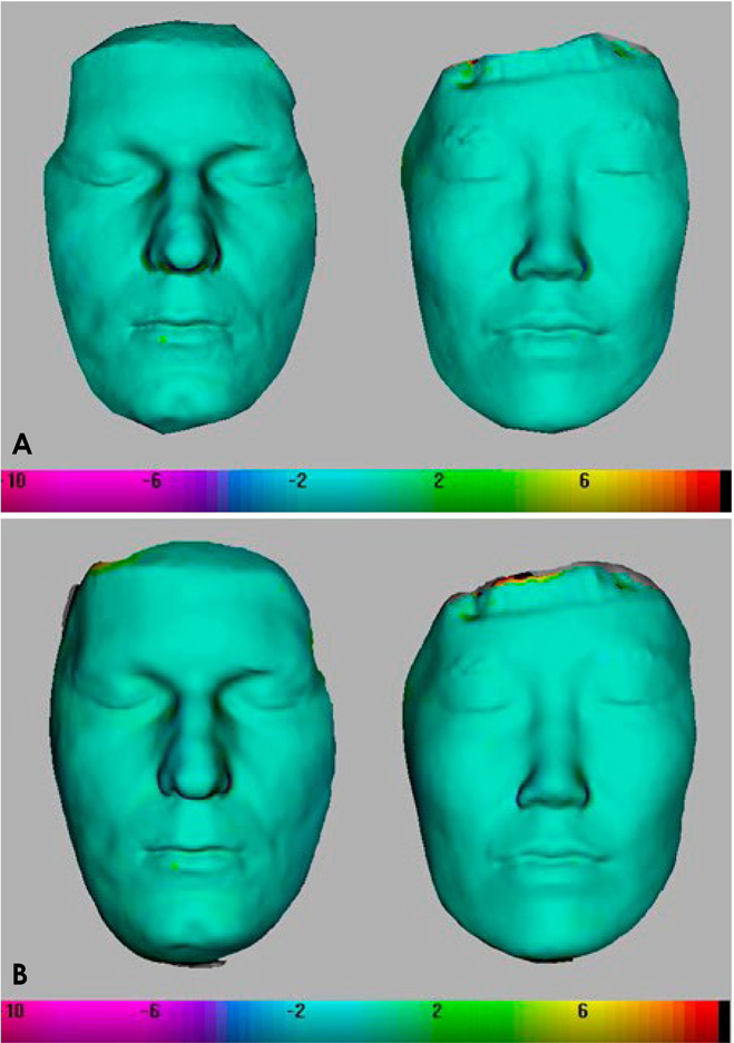
Fig. 4 Colour maps showing the minimal surface differences between the reference computed tomography scans and (A) single-capture Vectra images and (B) merged Vectra images, for both facial casts.

Fig. 5 Colour maps of both casts showing the surface deviations between the single-capture Scanify and single-capture Vectra H1 images in millimetres. Each pixel point was allocated a numerical value denoting the distance between corresponding points on the two 3-dimensional images.
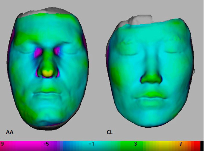
Fig. 6 Colour maps of both casts showing the surface differences between the single-capture Scanify and merged Vectra H1 images in millimetres.

Fig. 7 Colour maps of both casts showing the surface differences between the merged Scanify and merged Vectra H1 images in millimetres.
Reference
-
1. Choi JW, Lee JY, Oh TS, Kwon SM, Yang SJ, Koh KS. Frontal soft tissue analysis using a 3 dimensional camera following two-jaw rotational orthognathic surgery in skeletal class III patients. J Craniomaxillofac Surg. 2014; 42:220–226.
Article2. Verhoeven TJ, Coppen C, Barkhuysen R, Bronkhorst EM, Merkx MA, Bergé SJ, et al. Three dimensional evaluation of facial asymmetry after mandibular reconstruction: validation of a new method using stereophotogrammetry. Int J Oral Maxillofac Surg. 2013; 42:19–25.
Article3. Artopoulos A, Rosenberg R, Coward TJ. Three-dimensional imaging of the face using a portable, single camera, digital stereophotogrammetry device. In : Optical Measurement Techniques for Systems and Structures (OPTIMESS) Meeting; 2015 April 8-9; Antwerp, Belgium.4. Ozsoy U, Demirel BM, Yildirim FB, Tosun O, Sarikcioglu L. Method selection in craniofacial measurements: advantages and disadvantages of 3D digitization method. J Craniomaxillofac Surg. 2009; 37:285–290.
Article5. Plooij JM, Swennen GR, Rangel FA, Maal TJ, Schutyser FA, Bronkhorst EM, et al. Evaluation of reproducibility and reliability of 3D soft tissue analysis using 3D stereophotogrammetry. Int J Oral Maxillofac Surg. 2009; 38:267–273.
Article6. van Loon B, Maal TJ, Plooij JM, Ingels KJ, Borstlap WA, Kuijpers-Jagtman AM, et al. 3D stereophotogrammetric assessment of pre- and postoperative volumetric changes in the cleft lip and palate nose. Int J Oral Maxillofac Surg. 2010; 39:534–540.
Article7. Farkas LG. Anthropometry of the head and face. 2nd ed. New York: Raven Press;1994.8. Gwilliam JR, Cunningham SJ, Hutton T. Reproducibility of soft tissue landmarks on three-dimensional facial scans. Eur J Orthod. 2006; 28:408–415.
Article9. Krimmel M, Kluba S, Dietz K, Reinert S. Assessment of precision and accuracy of digital surface photogrammetry with the DSP 400 system. Biomed Tech (Berl). 2005; 503:45–53.10. Maal TJ, Verhamme LM, van Loon B, Plooij JM, Rangel FA, Kho A, et al. Variation of the face in rest using 3D stereophotogrammetry. Int J Oral Maxillofac Surg. 2011; 40:1252–1257.
Article11. Metzger MC, Hohlweg-Majert B, Schön R, Teschner M, Gellrich NC, Schmelzeisen R, et al. Verification of clinical precision after computer-aided reconstruction in craniomaxillofacial surgery. Oral Surg Oral Med Oral Pathol Oral Radiol Endod. 2007; 104:e1–e10.
Article12. Swennen GR, Schutyser F, Lemaitre A, Malevez C, De Mey A. Accuracy and reliability of 3-D CT versus 3-D stereo photogrammetry based facial soft tissue analysis. Int J Oral Maxillofac Surg. 2005; 34:73. (Abstr.).
Article13. Hajeer MY, Ayoub AF, Millett DT, Bock M, Siebert JP. Three-dimensional imaging in orthognathic surgery: the clinical application of a new method. Int J Adult Orthodon Orthognath Surg. 2002; 17:318–330.14. Germeç-Çakan D, Canter HI, Nur B, Arun T. Comparison of facial soft tissue measurements on three-dimensional images and models obtained with different methods. J Craniofac Surg. 2010; 21:1393–1399.15. Phillips C, Greer J, Vig P, Matteson S. Photocephalometry: errors of projection and landmark location. Am J Orthod. 1984; 86:233–243.
Article
- Full Text Links
-
- Actions
-
Cited
- CITED
-
- Close
- Share
-
- Similar articles
-
- Portable Low-Cost MRI System Based on Permanent Magnets/Magnet Arrays
- Use of an Extraoral Transfer Jig and a Handheld Face Scanner App for Integrating Face Scan Data into Prosthesis Design
- 3D Facial Scanners: How to Make the Right Choice for Orthodontists
- Feasibility Study of Utilizing a Portable Spectrophotometer to Assess the Radiation Dose Delivered to the Radiochromic Film
- Usefulness of the Portable Cystometer
