J Rheum Dis.
2019 Jan;26(1):83-84. 10.4078/jrd.2019.26.1.83.
Eosinophilic Granulomatosis with Polyangiitis Diagnosed by Gallbladder Tissue
- Affiliations
-
- 1Department of Internal Medicine, Jeju National University School of Medicine, Jeju, Korea. slera@yahoo.com
- 2Department of Pathology, Jeju National University School of Medicine, Jeju, Korea.
- KMID: 2442039
- DOI: http://doi.org/10.4078/jrd.2019.26.1.83
Abstract
- No abstract available.
Figure
-

Figure 1. Abdominal and pelvic computed tomography scan showing diffuse wall thickening of the gallbladder with some irregularity.
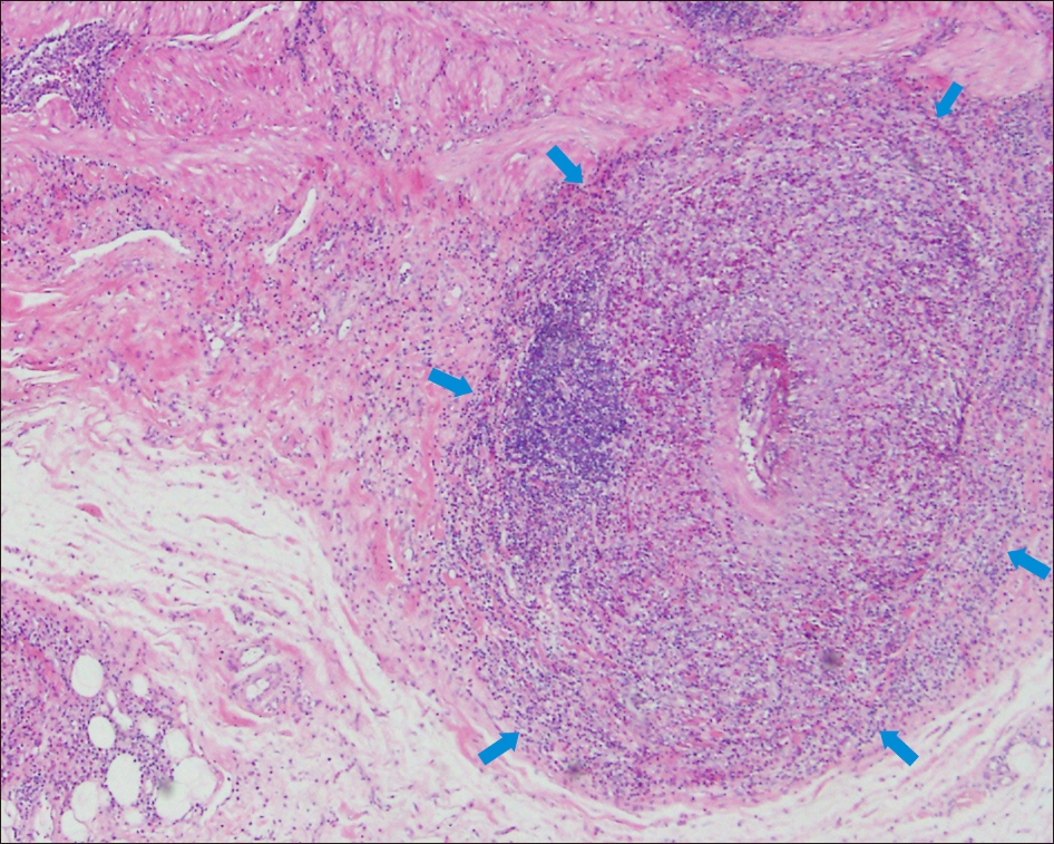
Figure 2. Granuloma surrounds the blood vessel (blue arrows) and granulomatous inflammation is also noted in and around the blood vessels (H&E, ×40).
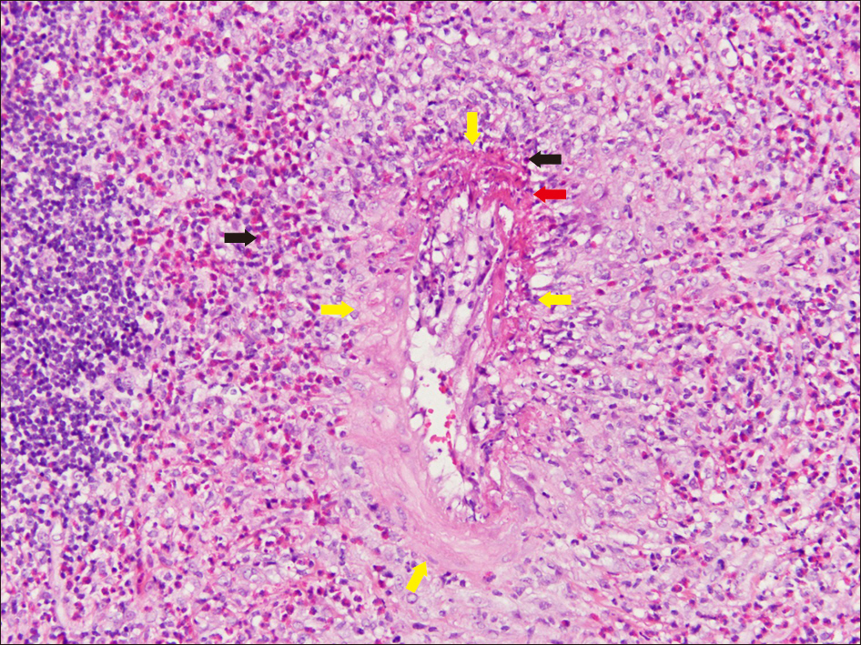
Figure 3. The vessel wall (yellow arrows) is destructed by inflammatory infiltrates in the right upper area of the vessel (vasculitis with fibrinoid necrosis, red arrow). Many eosinophils which have bright red colored cytoplasm infiltrates into and around the vessel (black arrows) (H&E, ×200).
Reference
- Full Text Links
-
- Actions
-
Cited
- CITED
-
- Close
- Share
-
- Similar articles
-
- Eosinophilic Annular Erythema in a Patient with Eosinophilic Granulomatosis with Polyangiitis (Churg-Strauss Syndrome)
- Granulomatosis with Polyangiitis Presented with Temporal Headache and Tenderness
- A Case of Eosinophilic Granulomatosis with Polyangiitis with a Clinical Presentation Similar to Erythema Gyratum Repens
- Eosinophilic granulomatosis with polyangiitis presenting as an endobronchial nodule and atelectasis: A case report
- A Retrospective Analysis of Granulomatosis with Polyangiitis with Ocular Manifestations
