Ann Dermatol.
2019 Apr;31(2):235-237. 10.5021/ad.2019.31.2.235.
A Case of Unusual Juvenile Xanthogranuloma on the Subungual Area
- Affiliations
-
- 1Department of Dermatology, Samsung Medical Center, Sungkyunkwan University School of Medicine, Seoul, Korea. dylee@skku.edu
- KMID: 2439076
- DOI: http://doi.org/10.5021/ad.2019.31.2.235
Abstract
- No abstract available.
MeSH Terms
Figure
-
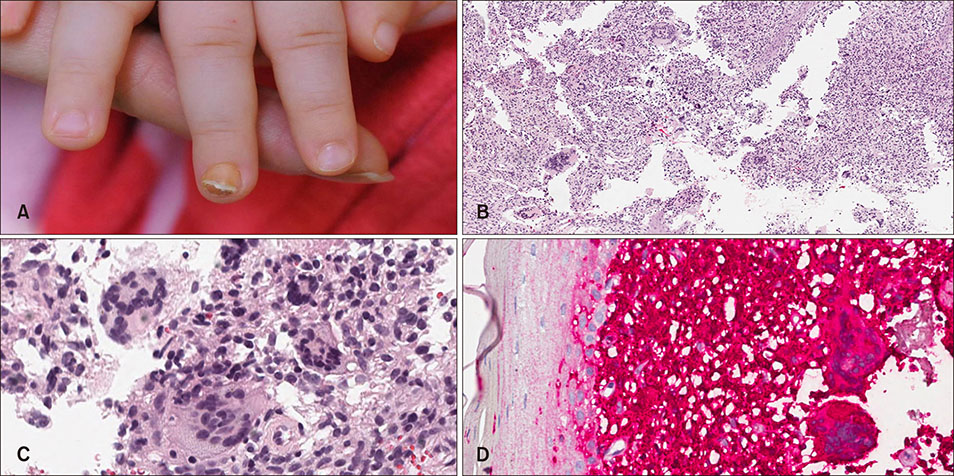
Fig. 1 (A) Yellow firm nodule under the nail right 3rd finger nail plate. (B) Dense intradermal histiocytic infiltrates and a number of giant cells (H&E, ×200). (C) Foreign body giant cells with haphazard nuclear arrangement were mainly observed (H&E, ×400). (D) All of histiocytes were positive for CD163 (CD163, ×400).
Reference
-
1. Frumkin A, Roytman M, Johnson SF. Juvenile xanthogranuloma underneath a toenail. Cutis. 1987; 40:244–245.2. Chang P, Baran R, Villanueva C, Samayoa M, Perrin C. Juvenile xanthogranuloma beneath a fingernail. Cutis. 1996; 58:173–174.3. Piraccini BM, Fanti PA, Iorizzo M, Tosti A. Juvenile xanthogranuloma of the proximal nail fold. Pediatr Dermatol. 2003; 20:307–308.
Article4. Kim EJ, Kim MY, Kim HO, Park YM. Juvenile xanthogranuloma of the finger: an unusual localization. J Dermatol. 2007; 34:590–592.
Article5. Kim JK, Kim B, Won CH, Chang SE, Lee MW, Choi JH, et al. Subungual juvenile xanthogranuloma. Korean J Dermatol. 2012; 50:354–357.
