Corneoconjunctival manifestations of lymphoma in three dogs
- Affiliations
-
- 1Department of Veterinary Clinical Sciences, College of Veterinary Medicine and Research Institute for Veterinary Science, Seoul National University, Seoul 08826, Korea. kmseo@snu.ac.kr
- 2I Animal Medical Center, Bucheon 14427, Korea.
- KMID: 2434785
- DOI: http://doi.org/10.4142/jvs.2019.20.1.98
Abstract
- An 8-year-old Shih Tzu, a 5-year-old Maltese, and a 10-year-old Maltese presented with conjunctival hyperemia and peripheral corneal edema. Severe conjunctival thickening with varying degrees of corneal extension was observed. Cytological examination showed many large lymphocytes with malignant changes in the conjunctiva which was consistent with findings in fine-needle aspiration samples taken from regional lymph nodes. They were diagnosed as having Stage V multicentric lymphoma. When conjunctival thickening is observed in canine patients with multicentric lymphoma, conjunctival metastasis with infiltration of neoplastic lymphoid cells should be included in the differential diagnosis.
Keyword
MeSH Terms
Figure
-
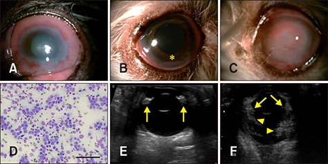
Fig. 1 Clinical appearance on initial presentation and the diagnostic approaches used. (A) The left eye in C1 (an 8-year-old neutered male Shih Tzu). (B) The right eye in C2 (a 5-year-old neutered male Maltese). (C) The left eye in C3 (a 10-year-old neutered male Maltese). Conjunctival thickening and hyperemia with corneal perilimbal (C1 and C2) or generalized (C3) edema and neovascularization were observed. Hypopyon (asterisk) was also seen in C2 (B). (D) The cytologic appearance of a fine-needle aspirate from conjunctiva in C1. Medium-sized to large-sized lymphocytes with malignant changes were detected. 400×. Scale bar = 100 µm. (E and F) Respective B-scan ultrasound images for C1 and C3. Thickening of the ciliary body (arrows) and echogenic masses within the vitreous body (arrowheads) with vitreous degeneration were evident.
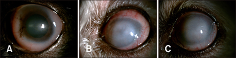
Fig. 2 Clinical appearance on follow-up examination. (A) The left eye in C1 (an 8-year-old neutered male Shih Tzu) after one month; the perilimbal corneal involvement improved and the conjunctival thickening and hyperemia also subsided. (B and C) The left eye in C3 (a 10-year-old neutered male Maltese) after 3 and 7 weeks, respectively; the conjunctival thickening subsided and the corneal edema and neovascularization also decreased. The corneoconjunctival lesions improved over time despite persistent corneal fibrosis.
Reference
-
1. Comazzi S, Gelain ME. Use of flow cytometric immunophenotyping to refine the cytological diagnosis of canine lymphoma. Vet J. 2011; 188:149–155.
Article2. Ettinger SN. Principles of treatment for canine lymphoma. Clin Tech Small Anim Pract. 2003; 18:92–97.
Article3. Hendrix DVH. Diseases and surgery of the canine anterior uvea. In : Gelatt KN, Gilger BC, Kern TJ, editors. Veterinary Ophthalmology. 5th ed. Ames: Wiley-Blackwell;2013. p. 1146–1198.4. Krohne SG, Henderson NM, Richardson RC, Vestre WA. Prevalence of ocular involvement in dogs with multicentric lymphoma: prospective evaluation of 94 cases. Vet Comp Ophthalmol. 1994; 4:127–135.5. Lanza MR, Musciano AR, Dubielzig RD, Durham AC. Clinical and pathological classification of canine intraocular lymphoma. Vet Ophthalmol. 2018; 21:167–173.
Article6. Ledbetter EC, Gilger BC. Diseases and surgery of the canine cornea and sclera. In : Gelatt KN, Gilger BC, Kern TJ, editors. Veterinary Ophthalmology. 5th ed. Ames: Wiley-Blackwell;2013. p. 976–1049.7. Maggs DJ. Conjunctiva. In : Maggs DJ, Miller PE, Ofri R, editors. Slatter's Fundamentals of Veterinary Ophthalmology. 5th ed. St. Louis: Elsevier Saunders;2013. p. 140–154.8. Olbertz L, Lima L, Langohr I, Werner J, Teixeira L, Montiani-Ferreira F. Supposed primary conjunctival lymphoma in a dog. Vet Ophthalmol. 2013; 16:Suppl 1. 100–104.
Article9. Ota-Kuroki J, Ragsdale JM, Bawa B, Wakamatsu N, Kuroki K. Intraocular and periocular lymphoma in dogs and cats: a retrospective review of 21 cases (2001-2012). Vet Ophthalmol. 2014; 17:389–396.
Article10. Stewart CJ, Duncan JA, Farquharson M, Richmond J. Fine needle aspiration cytology diagnosis of malignant lymphoma and reactive lymphoid hyperplasia. J Clin Pathol. 1998; 51:197–203.
Article11. Swanson JF. Ocular manifestations of systemic disease in the dog and cat: recent developments. Vet Clin North Am Small Anim Pract. 1990; 20:849–867.12. Vail DM, Dobson JM. Tumours of the haemopoietic system and spleen. In : Dobson JM, Lascelles BDX, editors. BSAVA Manual of Canine and Feline Oncology. 3rd ed. Gloucester: British Small Animal Veterinary Association;2010. p. 285–308.13. Vail DM, Pinkerton ME, Young KM. Hematopoietic tumors. In : Withrow SJ, Vail DM, Page RL, editors. Withrow and MacEwen's Small Animal Clinical Oncology. 5th ed. Philadelphia: Saunders;2013. p. 608–678.14. Vascellari M, Multari D, Mutinelli F. Unicentric extranodal lymphoma of the upper eyelid conjunctiva in a dog. Vet Ophthalmol. 2005; 8:67–70.
Article
- Full Text Links
-
- Actions
-
Cited
- CITED
-
- Close
- Share
-
- Similar articles
-
- Manifestations of lymphoma in plain chest x-ray
- A Case of Non-Hodgkin's Lymphoma of the Oral Cavity Presenting as a Buccal Mass
- A Rare Case of Intestinal T-cell Lymphoma with Multiple Complications
- Pulmonary Involvement of T-cell type Lymphoma with Rapid, Bilateral Infiltration and High Fever Simulating Pueumonia
- Primary Lymphoma of Cervix
