Automated Detection of Retinal Nerve Fiber Layer by Texture-Based Analysis for Glaucoma Evaluation
- Affiliations
-
- 1Department of Computer Science, Faculty of Computer Science and Information Technology, Mulawarman University, Samarinda, Indonesia. anindita@unmul.ac.id
- 2Department of Computer Science and Electronics, Faculty of Mathematics and Natural Sciences, Universitas Gadjah Mada, Yogyakarta, Indonesia.
- 3Faculty of Medicine, Universitas Gadjah Mada, Yogyakarta, Indonesia.
- KMID: 2434549
- DOI: http://doi.org/10.4258/hir.2018.24.4.335
Abstract
OBJECTIVES
The retinal nerve fiber layer (RNFL) is a site of glaucomatous optic neuropathy whose early changes need to be detected because glaucoma is one of the most common causes of blindness. This paper proposes an automated RNFL detection method based on the texture feature by forming a co-occurrence matrix and a backpropagation neural network as the classifier.
METHODS
We propose two texture features, namely, correlation and autocorrelation based on a co-occurrence matrix. Those features are selected by using a correlation feature selection method. Then the backpropagation neural network is applied as the classifier to implement RNFL detection in a retinal fundus image.
RESULTS
We used 40 retinal fundus images as testing data and 160 sub-images (80 showing a normal RNFL and 80 showing RNFL loss) as training data to evaluate the performance of our proposed method. Overall, this work achieved an accuracy of 94.52%.
CONCLUSIONS
Our results demonstrated that the proposed method achieved a high accuracy, which indicates good performance.
Keyword
MeSH Terms
Figure
-
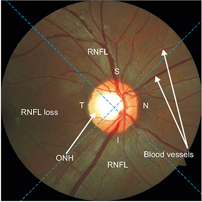
Figure 1 Overview of the retinal structure in retinal fundus image of the right eye and of the sector partition. RNFL: retinal nerve fiber layer, ONH: optic nerve head.

Figure 2 Stage diagram of the proposed retinal nerve fiber layer (RNFL) detection method. ROI: region of interest, ONH: optic nerve head.

Figure 3 Original image and the results of region of interest (ROI) detection: (A) retinal fundus image, (B) boundary of optic nerve head (ONH), (C) updated position of ONH, and (D) ROI image with sector division.
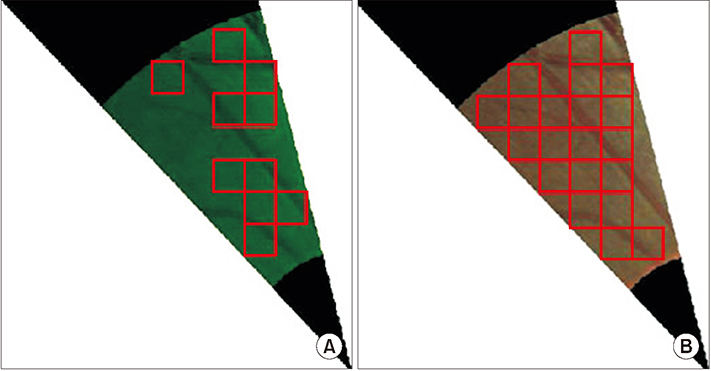
Figure 4 Examples of classification results on each patch in a sub-sector with red-free (A) and RGB (B) images.
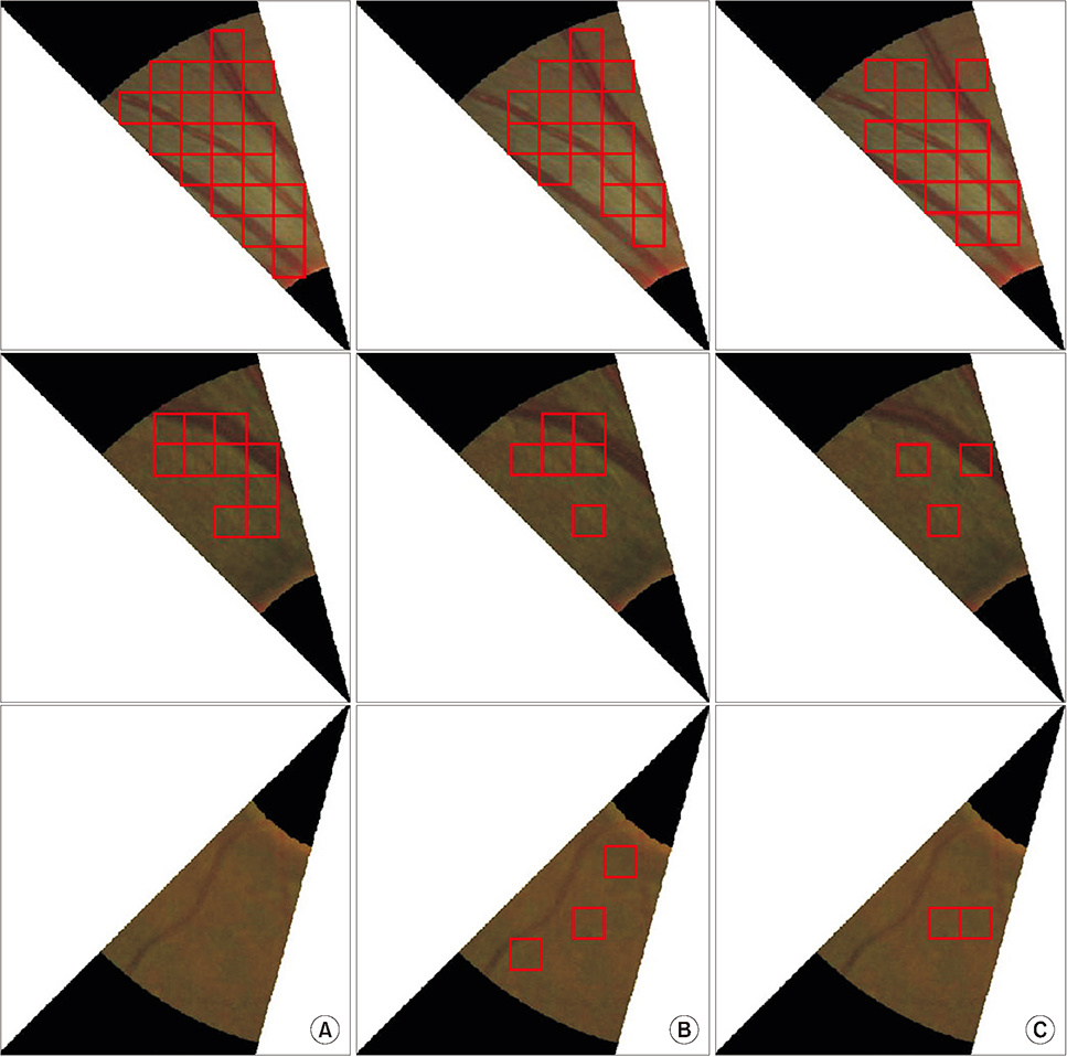
Figure 5 Comparison of retinal nerve fiber layer detection result obtained using three different classifiers: (A) backpropagation neural network, (B) support vector machine, and (C) k-nearest neighbor.

Figure 6 Partition of sectors into sub-sectors and the ground truth. RNFL: retinal nerve fiber layer.
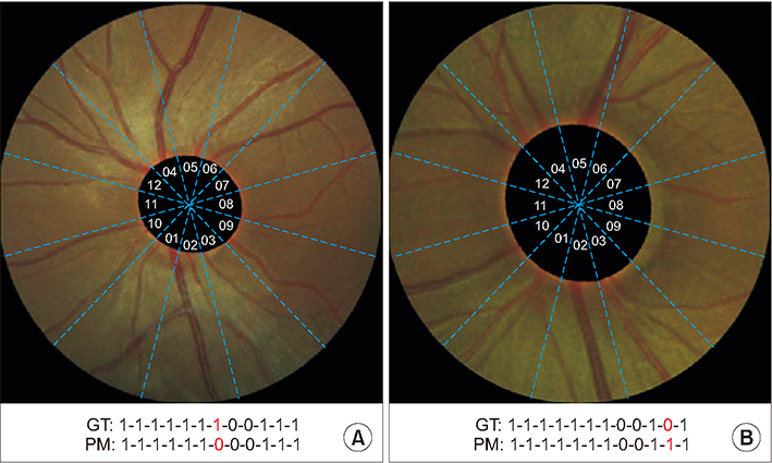
Figure 7 Examples of erroneous retinal nerve fiber layer detection results obtained by the proposed method: (A) false negative and (B) false positive. GT: ground truth, PM: proposed method.
Cited by 1 articles
-
Automatic Method for Optic Disc Segmentation Using Deep Learning on Retinal Fundus Images
Anindita Septiarini, Hamdani Hamdani, Emy Setyaningsih, Eko Junirianto, Fitri Utaminingrum
Healthc Inform Res. 2023;29(2):145-151. doi: 10.4258/hir.2023.29.2.145.
Reference
-
1. Hollo G. The optic nerve in glaucoma. In : Choplin NT, Traverso CE, editors. Atlas of glaucoma. 3rd ed. Boca Raton (FL): Taylor & Francis;2014. p. 61–71.2. Chakrabarty L, Joshi GD, Chakravarty A, Raman GV, Krishnadas SR, Sivaswamy J. Automated detection of glaucoma from topographic features of the optic nerve head in color fundus photographs. J Glaucoma. 2016; 25(7):590–597.
Article3. Quigley HA. Number of people with glaucoma worldwide. Br J Ophthalmol. 1996; 80(5):389–393.
Article4. Quigley HA, Broman AT. The number of people with glaucoma worldwide in 2010 and 2020. Br J Ophthalmol. 2006; 90(3):262–267.
Article5. Leung CK. Detecting optic nerve head deformation and retinal nerve fiber layer thinning in glaucoma progression. Taiwan J Ophthalmol. 2015; 5(2):50–55.
Article6. Besenczi R, Toth J, Hajdu A. A review on automatic analysis techniques for color fundus photographs. Comput Struct Biotechnol J. 2016; 14:371–384.
Article7. Fondon I, Nunez F, Tirado M, Jimenez S, Alemany P, Abbas Q, et al. Automatic cup-to-disc ratio estimation using active contours and color clustering in fundus images for glaucoma diagnosis. In : Campilho A, Kamel M, editors. Image analysis and recognition. Heidelberg: Springer;2012. p. 390–399.8. Hsiao HK, Liu CC, Yu CY, Kuo SW, Yu SS. A novel optic disc detection scheme on retinal images. Expert Syst Appl. 2012; 39(12):10600–10606.
Article9. Septiarini A, Harjoko A, Pulungan R, Ekantini R. Optic disc and cup segmentation by automatic thresholding with morphological operation for glaucoma evaluation. Signal Image Video Process. 2017; 11(5):945–952.
Article10. Pourreza-Shahri R, Tavakoli M, Kehtarnavaz N. Computationally efficient optic nerve head detection in retinal fundus images. Biomed Signal Process Control. 2014; 11:63–73.
Article11. Singh A, Dutta MK, ParthaSarathi M, Uher V, Burget R. Image processing based automatic diagnosis of glaucoma using wavelet features of segmented optic disc from fundus image. Comput Methods Programs Biomed. 2016; 124:108–120.
Article12. Issac A, Partha Sarathi M, Dutta MK. An adaptive threshold based image processing technique for improved glaucoma detection and classification. Comput Methods Programs Biomed. 2015; 122(2):229–244.
Article13. Marin D, Gegundez-Arias ME, Suero A, Bravo JM. Obtaining optic disc center and pixel region by automatic thresholding methods on morphologically processed fundus images. Comput Methods Programs Biomed. 2015; 118(2):173–185.
Article14. Mary MC, Rajsingh EB, Jacob JK, Anandhi D, Amato U, Selvan SE. An empirical study on optic disc segmentation using an active contour model. Biomed Signal Process Control. 2015; 18:19–29.
Article15. Mittapalli PS, Kande GB. Segmentation of optic disk and optic cup from digital fundus images for the assessment of glaucoma. Biomed Signal Process Control. 2016; 24:34–46.
Article16. Cheng J, Liu J, Xu Y, Yin F, Wong DW, Tan NM, et al. Superpixel classification based optic disc and optic cup segmentation for glaucoma screening. IEEE Trans Med Imaging. 2013; 32(6):1019–1032.
Article17. Dashtbozorg B, Mendonca AM, Campilho A. Optic disc segmentation using the sliding band filter. Comput Biol Med. 2015; 56:1–12.
Article18. Morales S, Naranjo V, Angulo U, Alcaniz M. Automatic detection of optic disc based on PCA and mathematical morphology. IEEE Trans Med Imaging. 2013; 32(4):786–796.
Article19. FGazarek J, Jan J, Kolar R, Odstrcilik J. Retinal nerve fibre layer detection in fundus camera images compared to results from optical coherence tomography. In : Proceedings of 2011 International Conference on Image Information Processing (ICIIP); 2011 Nov 3-5; Shimla, India. p. 1–5.20. Odstrcilik J, Kolar R, Jan J, Gazarek J, Kuna Z, Vodakova M. Analysis of retinal nerve fiber layer via Markov random fields in color fundus images. In : Proceedings of 2012 19th International Conference on Systems, Signals and Image Processing (IWSSIP); 2012 Apr 11-13; Vienna, Austria. p. 504–507.21. Prageeth PG, David J, Kumar AS. Early detection of retinal nerve fiber layer defects using fundus image processing. In : Proceedings of 2011 IEEE Recent Advances in Intelligent Computational Systems (RAICS); 2011 Sep 22-24; Trivandrum, India. p. 930–936.22. Kolar R, Jan J. Detection of glaucomatous eye via color fundus images using fractal dimensions. Radioeng. 2008; 17(3):109–114.23. Joshi GD, Sivaswamy J, Prashanth R, Krishnadas SR. Detection of peri-papillary atrophy and RNFL defect from retinal images. In : Campilho A, Kamel M, editors. Image Analysis and Recognition. Heidelberg: Springer;2002. p. 400–407.24. Soh LK, Tsatsoulis C. Texture analysis of SAR sea ice imagery using gray level co-occurrence matrices. IEEE Trans Geosci Remote Sens. 1999; 37(2):780–795.
Article25. Kim TY, Son J, Kim KG. The recent progress in quantitative medical image analysis for computer aided diagnosis systems. Healthc Inform Res. 2011; 17(3):143–149.
Article26. Septiarini A, Harjoko A, Pulungan R, Ekantini R. Automatic detection of peripapillary atrophy in retinal fundus images using statistical features. Biomed Signal Process Control. 2018; 45:151–159.
Article27. Septiarini A, Khairina DM, Kridalaksana AH, Hamdani H. Automatic glaucoma detection method applying a statistical approach to fundus images. Healthc Inform Res. 2018; 24(1):53–60.
Article28. Yousefi S, Goldbaum MH, Balasubramanian M, Jung TP, Weinreb RN, Medeiros FA, et al. Glaucoma progression detection using structural retinal nerve fiber layer measurements and functional visual field points. IEEE Trans Biomed Eng. 2014; 61(4):1143–1154.
Article29. Orlando JI, Prokofyeva E, Del Fresno M, Blaschko MB. An ensemble deep learning based approach for red lesion detection in fundus images. Comput Methods Programs Biomed. 2018; 153:115–127.
Article30. Raghavendra U, Fujita H, Bhandary SV, Gudigar A, Tan JH, Acharya UR. Deep convolution neural network for accurate diagnosis of glaucoma using digital fundus images. Inf Sci. 2018; 441:41–49.
Article
- Full Text Links
-
- Actions
-
Cited
- CITED
-
- Close
- Share
-
- Similar articles
-
- Reproducibility of Retinal Nerve Fiber Layer Thickness Evaluation by Nerve Fiber Analyzer
- Biometry of Retinal Nerve Fiber Layer Thickness by NFA
- Early Detection of Glaucoma with Retinal Nerve Fiber Layer Photograph
- Influence of Diabetes Mellitus on the Retinal Ne rve Fiber Layer Thickness Measurement by Nerve Fiber Analyzer
- Clinical Evaluation of Unilateral Open-Angle Glaucoma: A Two-Year Follow-Up Study
