Korean J Ophthalmol.
2019 Feb;33(1):99-100. 10.3341/kjo.2018.0054.
Atypical Pattern of Choroidal Hypopigmentation with Cutaneous Vitiligo
- Affiliations
-
- 1Department of Ophthalmology and Visual Science, Seoul St. Mary's Hospital, The Catholic University of Korea College of Medicine, Seoul, Korea. parkyh@catholic.ac.kr
- 2Department of Ophthalmology and Visual Science, St. Vincent's Hospital, The Catholic University of Korea College of Medicine, Suwon, Korea.
- KMID: 2434312
- DOI: http://doi.org/10.3341/kjo.2018.0054
Abstract
- No abstract available.
MeSH Terms
Figure
-
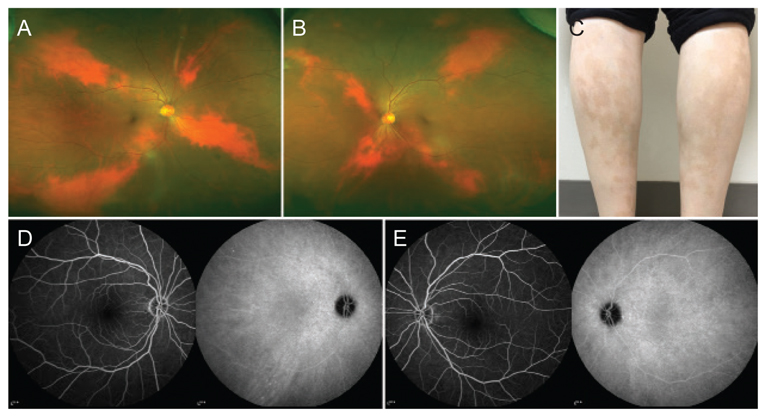
Fig. 1 Initial fundus photography of (A) the right eye and (B) left eye, and X-shaped areas of choroidal hypopigmentation were observed. (C) The patient reported having cutaneous vitiligo of the lower extremities. (D,E) Tiny scattered hypofluorescent spots were present on f luorescein angiography and hyperf luorescent spots were observed on indocyanine green angiography bilaterally. Informed consent for medical photographs was obtained.
Reference
-
1. Biswas G, Barbhuiya JN, Biswas MC, et al. Clinical pattern of ocular manifestations in vitiligo. J Indian Med Assoc. 2003; 101:478–480.2. Elder DE. Lever's histopathology of the skin. Philadelphia: Lippincott Williams & Wilkins;2014. p. 260–268.3. Prignano F, Betts CM, Lotti T. Vogt-Koyanagi-Harada disease and vitiligo: where does the illness begin? J Electron Microsc (Tokyo). 2008; 57:25–31.
Article4. Shields CL, Ramasubramanian A, Kunz WB, et al. Choroidal vitiligo masquerading as large choroidal nevus: a report of four cases. Ophthalmology. 2010; 117:109–113.
Article
- Full Text Links
-
- Actions
-
Cited
- CITED
-
- Close
- Share
-
- Similar articles
-
- A Case of Hypopigmented Mycosis Fungoides
- Immunology of Vitiligo
- A Case of Hypopigmented Mycosis Fungoides
- Halo-Like Disappearance of Café au Lait Spot: A Clue for the Role of Autoimmunity and Somatic Mosaicism in Segmental Vitiligo
- Hypopigmentary Disorders Excluding Vitiligo : Clinical Features in 301 Patients
