Palpebral Conjunctival and Tarsal Stromal Tumor
- Affiliations
-
- 1Department of Ophthalmology, Sanggye Paik Hospital, Inje University College of Medicine, Seoul, Korea.
- 2ROK II Corps 702 Special Assault Regiment, Hwacheon, Korea.
- 3Department of Ophthalmology, Sanggye Paik Hospital, Inje University College of Medicine, Seoul, Korea.
- 4Department of Pathology, Sanggye Paik Hospital, Inje University College of Medicine, Seoul, Korea.
- 5Department of Ophthalmology, Sanggye Paik Hospital, Inje University College of Medicine, Seoul, Korea. sylvia8@hanmail.net
- KMID: 2434311
- DOI: http://doi.org/10.3341/kjo.2018.0059
Abstract
- No abstract available.
Figure
-
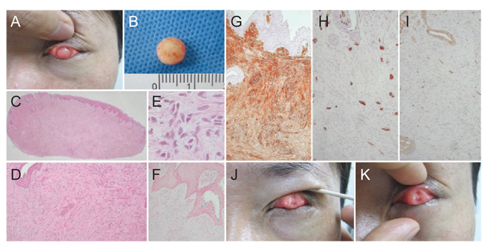
Fig. 1 (A) Clinical appearance of the mass: The mass presented on the left upper palpebral conjunctiva, showing reddish and firm characteristics. (B) Shave biopsy of the mass: The mass was dissected with shave biopsy and was 8 mm in diameter. (C) A nodular mass measuring 0.8 cm in its greatest dimension with tenting squamo-columnar epithelium was noted (H&E, ×10). (D) The mass was composed of interlacing spindle cells with scanty myxoid stroma (H&E, ×100). (E) The tumor cells disclosed mild to moderate cellularity and mild nuclear pleomorphism. Only one mitosis was identified in the whole field (inlet) (H&E, ×400). (F) Alcian blue special staining revealed scanty amount of stromal mucin in contrast to the cytoplasmic mucin of the intraepithelial goblet cells (×100). (G) Immunohistochemical staining of tumor cells disclosed diffuse and strong positivity for CD34 (×100). (H) The immunohistochemical stains were negative for smooth muscle actin (×100). (I) C-kit immunohistochemical staining was only reactive in mast cells (×100). Written informed conset was obtained from the patient for publication of this case report and any accompanying images. (J) A remnant mass 5 mm in diameter was noticeable. (K) Clinical appearance of the patient 2 months after the shave biopsy: proliferation of the remnant mass was found, and the mass measured 6 mm in diameter.
Reference
-
1. Albert DM, Miller JW, Azar DT, Blodi BA. Albert and Jakobiec's principles and practice of ophthalmology. 3rd ed. Amsterdam: Elsevier;2008. p. 46–48.2. Herwig MC, Wells JR, Grossniklaus HE. Conjunctival stromal tumor: report of 4 cases. Ophthalmology. 2012; 119:682–687.
Article3. Kumar V, Abbas AK, Fausto N, et al. Robbins and Cotran pathologic basis of disease. 8th ed. Philadelphia: Saunders;2010. p. 1319–1343.4. Manganoni AM, Pavoni L, Gualdi G, et al. Dermatofibrosarcoma protuberans in an adolescent: a case report and review of the literature. J Pediatr Hematol Oncol. 2013; 35:383–387.5. Kong LQ, Zhu XD, Xu HX, et al. The clinical significance of the CD163+ and CD68+ macrophages in patients with hepatocellular carcinoma. PLoS One. 2013; 8:e59771.
Article
- Full Text Links
-
- Actions
-
Cited
- CITED
-
- Close
- Share
-
- Similar articles
-
- Blepharoptosis Secondary to Local Conjunctival and Tarsal Amyloidosis
- Effects of Medpor(R) Sheet as Substitute for Tarsus in Eyelid Reconstruction
- Unilateral Eyelid Swelling Secondary to Local Palpebral Conjunctival Amyloidosis in a Young Patient
- A Conjunctival Myxoid Stromal Tumor
- Transconjunctival Lower Eyelid Widening using Tucking Method between Lower Eyelid Retractor and Lower Tarsal Plate
