Evaluation of Subconjunctival Remnant Particles after High-frequency Radio-wave Electrosurgery for Conjunctivochalasis
- Affiliations
-
- 1Department of Ophthalmology, Kangbuk Samsung Hospital, Sungkyunkwan University School of Medicine, Seoul, Korea. sashimi0@naver.com
- KMID: 2434300
- DOI: http://doi.org/10.3341/kjo.2018.0093
Abstract
- PURPOSE
To investigate the clinical manifestations and properties of remnant particles in the subconjunctival space after high-frequency radio-wave electrosurgery for conjunctivochalasis.
METHODS
We performed a retrospective, observational case series with in vitro experimental imaging in nine eyes from eight patients who presented with small dark-gray lesions during follow-up after high-frequency radio-wave electrosurgery for conjunctivochalasis. General examination including slit-lamp examination and visual acuity testing was performed preoperatively and postoperatively. During follow-up, we evaluated remnant particles and any other complications including granuloma or conjunctival injection with slit-lamp photography and anterior optical coherence tomography. Coagulation tips were investigated with scanning electron microscope and energy dispersive X-ray spectroscopy to analyze the insulating electrode and assess changes to tips after repeated use.
RESULTS
None of the patients included in this study experienced any change in visual acuity or major complications postoperatively. Small dark-gray lesions (0.3 to 0.5 mm in size) were observed in the inferior bulbar sub-conjunctival space in the location where high-frequency radio-wave electrosurgery had been performed. Cirrus high-definition optical coherence tomography images revealed focal hyper-reflection with a posterior shadow, suggesting foreign particles. Scanning electron microscopy and energy dispersive X-ray spectroscopy imaging analysis revealed peaks of carbon and fluorine complexes, consistent with the polytetrafluoroethylene coating on the electrode.
CONCLUSIONS
There were no instances of inflammatory reaction, particle migration, or major complications due to particles. Physicians should be aware of the possibility of remnant polytetrafluoroethylene particles in subconjunctival tissue when using insulated coagulation tips subjected to repeat sterilization.
Keyword
MeSH Terms
Figure
-
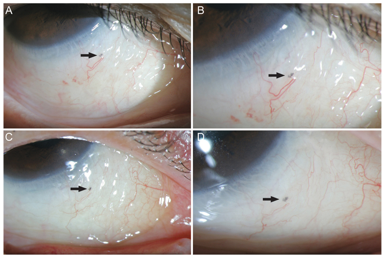
Fig. 1 Slit-lamp photographs of a dark-gray lesion at the inferior bulbar conjunctiva (arrow) observed in a 63-year-old woman. No observed changes were observed in the lesion 3 months after surgery: (A,B) 1 month postoperatively and (C,D) 3 months postoperatively.
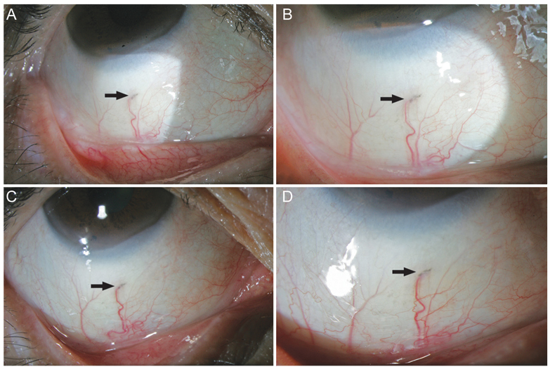
Fig. 2 Slit-lamp photographs of a dark-gray lesion at the inferior bulbar conjunctiva (arrow) observed in a 67-year-old woman. No observed changes were observed in the lesion 3 months after surgery: (A,B) 1 month postoperatively; (C,D) 3 months postoperatively.
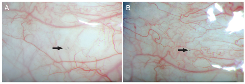
Fig. 3 Slit-lamp photographs of a 57-year-old man. One month after surgery, several tiny dark-gray lesions at nasal and temporal conjunctiva (arrow) were observed: 1 month postoperatively (A) at the nasal conjunctiva and (B) at the temporal conjunctiva.
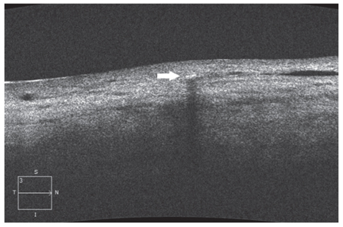
Fig. 4 High-definition optical coherence tomography images of a 63-year-old woman (right eye). Three months postoperatively, high-definition optical coherence tomography image showed a focal hyper-reflection with posterior shadowing (arrow) in the subconjunctival area.
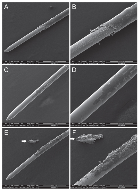
Fig. 5 Scanning electron microscopy images of the shaft of an insulated tip. (A,B) A new insulated fine-needle electrode. (C,D) A partially damaged insulated surface after a single use. (E,F) A severely damaged insulated surface after multiple uses. A large polytetrafluoroethylene particle peeling away from the shaft was noted (arrow) (scanning electron microscope, ×200, ×500).
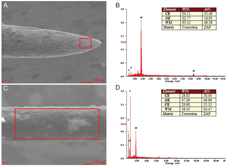
Fig. 6 Scanning electron microscopy (SEM) and energy-dispersive X-ray spectroscopy spectrum (EDS) images of the shaft of an insulated tip: (A) SEM image (×1,000) of the tip without insulation. (B) EDS image of the tip showing a prominent peak for wolframium (W) as well as other associated elements, including carbon (C) and oxygen (O). (C) SEM image (×1,000) of the insulated shaft. (D) EDS image of the shaft showing a prominent peak for fluorine (F) as well as for other associated elements, including C, O, and W. W = wolframium; C = carbon; O = oxygen; F = fluorine; Wt% = weight percent; At% = atomic percent; ZAF = ZAF corrections.
Reference
-
1. Hughes WL. Conjunctivochalasis. Am J Ophthalmol. 1942; 25:48–51.
Article2. Liu D. Conjunctivochalasis: a cause of tearing and its management. Ophthalmic Plast Reconstr Surg. 1986; 2:25–28.3. Meller D, Tseng SC. Conjunctivochalasis: literature review and possible pathophysiology. Surv Ophthalmol. 1998; 43:225–232.4. Jordan DR, Pelletier CR. Conjunctivochalasis. Can J Ophthalmol. 1996; 31:192–193.5. Meller D, Li DQ, Tseng SC. Regulation of collagenase, stromelysin, and gelatinase B in human conjunctival and conjunctivochalasis fibroblasts by interleukin-1beta and tumor necrosis factor-alpha. Invest Ophthalmol Vis Sci. 2000; 41:2922–2929.6. Otaka I, Kyu N. A new surgical technique for management of conjunctivochalasis. Am J Ophthalmol. 2000; 129:385–387.
Article7. Ahn JM, Choi CY, Seo KY. Surgical approach with high-frequency radiowave electrosurgery for superior limbic keratoconjunctivitis. Cornea. 2014; 33:210–214.
Article8. Han KE, Choi CY, Seo KY. Removal of lymphangiectasis using high-frequency radio wave electrosurgery. Cornea. 2013; 32:547–549.
Article9. Park J, Lee S, Suh E. Removal of conjunctival cyst with high-frequency radio-wave electrosurgery. Can J Ophthalmol. 2015; 50:378–383.
Article10. Woo KI, Choi CY. High-frequency radiowave electrosurgery for persistent conjunctival chemosis following cosmetic blepharoplasty. Plast Reconstr Surg. 2014; 133:1336–1342.
Article11. Youm DJ, Kim JM, Choi CY. Simple surgical approach with high-frequency radio-wave electrosurgery for conjunctivochalasis. Ophthalmology. 2010; 117:2129–2133.
Article12. Suh JS, Choi S. The effect of conjunctivochalasis surgery using a high-frequency radio-wave electrosurgical unit. J Korean Ophthalmol Soc. 2012; 53:1571–1576.
Article13. Lau C, Thibodeaux JR, Hanson RG, et al. Exposure to perfluorooctane sulfonate during pregnancy in rat and mouse. II: postnatal evaluation. Toxicol Sci. 2003; 74:382–392.
Article14. Seacat AM, Thomford PJ, Hansen KJ, et al. Subchronic toxicity studies on perfluorooctanesulfonate potassium salt in cynomolgus monkeys. Toxicol Sci. 2002; 68:249–264.
Article15. Johansson N, Fredriksson A, Eriksson P. Neonatal exposure to perfluorooctane sulfonate (PFOS) and perfluorooctanoic acid (PFOA) causes neurobehavioural defects in adult mice. Neurotoxicology. 2008; 29:160–169.
Article16. White SS, Kato K, Jia LT, et al. Effects of perfluorooctanoic acid on mouse mammary gland development and differentiation resulting from cross-foster and restricted gestational exposures. Reprod Toxicol. 2009; 27:289–298.
Article17. Bookstaff RC, Moore RW, Ingall GB, Peterson RE. Androgenic deficiency in male rats treated with perfluorodecanoic acid. Toxicol Appl Pharmacol. 1990; 104:322–333.
Article18. Nelson JW, Hatch EE, Webster TF. Exposure to polyfluoroalkyl chemicals and cholesterol, body weight, and insulin resistance in the general U.S. population. Environ Health Perspect. 2010; 118:197–202.
Article19. Sakr CJ, Kreckmann KH, Green JW, et al. Cross-sectional study of lipids and liver enzymes related to a serum biomarker of exposure (ammonium perfluorooctanoate or APFO) as part of a general health survey in a cohort of occupationally exposed workers. J Occup Environ Med. 2007; 49:1086–1096.
Article20. Steenland K, Tinker S, Frisbee S, et al. Association of perfluorooctanoic acid and perfluorooctane sulfonate with serum lipids among adults living near a chemical plant. Am J Epidemiol. 2009; 170:1268–1278.
Article21. Fairley KJ, Purdy R, Kearns S, et al. Exposure to the immunosuppressant, perf luorooctanoic acid, enhances the murine IgE and airway hyperreactivity response to ovalbumin. Toxicol Sci. 2007; 97:375–383.22. Yang Q, Xie Y, Alexson SE, et al. Involvement of the peroxisome proliferator-activated receptor alpha in the immunomodulation caused by peroxisome proliferators in mice. Biochem Pharmacol. 2002; 63:1893–1900.
Article23. Yang Q, Xie Y, Eriksson AM, et al. Further evidence for the involvement of inhibition of cell proliferation and development in thymic and splenic atrophy induced by the peroxisome proliferator perfluoroctanoic acid in mice. Biochem Pharmacol. 2001; 62:1133–1140.24. Alexander BH, Olsen GW, Burris JM, et al. Mortality of employees of a perfluorooctanesulphonyl fluoride manufacturing facility. Occup Environ Med. 2003; 60:722–729.
Article25. Gilliland FD, Mandel JS. Mortality among employees of a perf luorooctanoic acid production plant. J Occup Med. 1993; 35:950–954.26. Bloom MS, Kannan K, Spliethoff HM, et al. Exploratory assessment of perfluorinated compounds and human thyroid function. Physiol Behav. 2010; 99:240–245.
Article27. Dallaire R, Dewailly E, Pereg D, et al. Thyroid function and plasma concentrations of polyhalogenated compounds in Inuit adults. Environ Health Perspect. 2009; 117:1380–1386.
Article28. Olsen GW, Zobel LR. Assessment of lipid, hepatic, and thyroid parameters with serum perfluorooctanoate (PFOA) concentrations in fluorochemical production workers. Int Arch Occup Environ Health. 2007; 81:231–246.
Article29. Patrick CR. Thermal stability of polytetrafluoroethylene. Nature. 1958; 181:698.
Article30. Apelberg BJ, Witter FR, Herbstman JB, et al. Cord serum concentrations of perfluorooctane sulfonate (PFOS) and perfluorooctanoate (PFOA) in relation to weight and size at birth. Environ Health Perspect. 2007; 115:1670–1676.
Article31. Fei C, McLaughlin JK, Tarone RE, Olsen J. Perfluorinated chemicals and fetal growth: a study within the Danish National Birth Cohort. Environ Health Perspect. 2007; 115:1677–1682.
Article32. Washino N, Saijo Y, Sasaki S, et al. Correlations between prenatal exposure to perfluorinated chemicals and reduced fetal growth. Environ Health Perspect. 2009; 117:660–667.
Article
- Full Text Links
-
- Actions
-
Cited
- CITED
-
- Close
- Share
-
- Similar articles
-
- Removal of Eyelid Epidermal Cyst Using High-Frequency Radio-Wave Electrosurgery
- The Effect of Conjunctivochalasis Surgery Using a High-Frequency Radio-Wave Electrosurgical Unit
- Acute Exacerbation of Conjunctival Papilloma after High-frequency Radio Wave Electrosurgery for Conjunctivochalasis: A Case Report
- Two Cases of Corneal Neovascularization Treatment Using High-frequency Radio Wave Electrosurgery
- Treatment of Trichiasis and Districhiasis with Ellman Dento-Surg 90FFP
