Esthetic anterior restoration using 3M Lavaâ„¢ Esthetic monolithic zirconia
- Affiliations
-
- 1Department of Prosthodontics, College of Dentistry, Dankook University, Cheonan, Republic of Korea. syshin@dankook.ac.kr
- KMID: 2432367
- DOI: http://doi.org/10.14368/jdras.2018.34.4.306
Abstract
- Monolithic zirconia has been widely used in fixed partial dentures due to high strength and fracture toughness. Nevertheless, the usage of monolithic zirconia in anterior restoration was limited because of opacity. Recently, esthetic monolithic zirconia blocks are developed by improving translucency and using various shading systems. Manufacturer introduces 3M Lavaâ„¢ Esthetic with increased cubic phase and fluorescent ingredients is more esthetic than previous monolithic zirconia. This case report describes favorable anterior restorations using translucent monolithic zirconia.
MeSH Terms
Figure
-
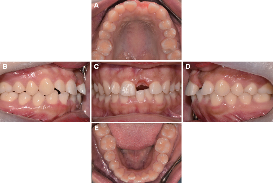
Fig. 1 Preoperative intraoral view showing crowding of mandibular anterior teeth. (A) Occlusal view of maxilla, (B) Right lateral view, (C) Frontal view, (D) Left lateral view, (E) Occlusal view of mandible.
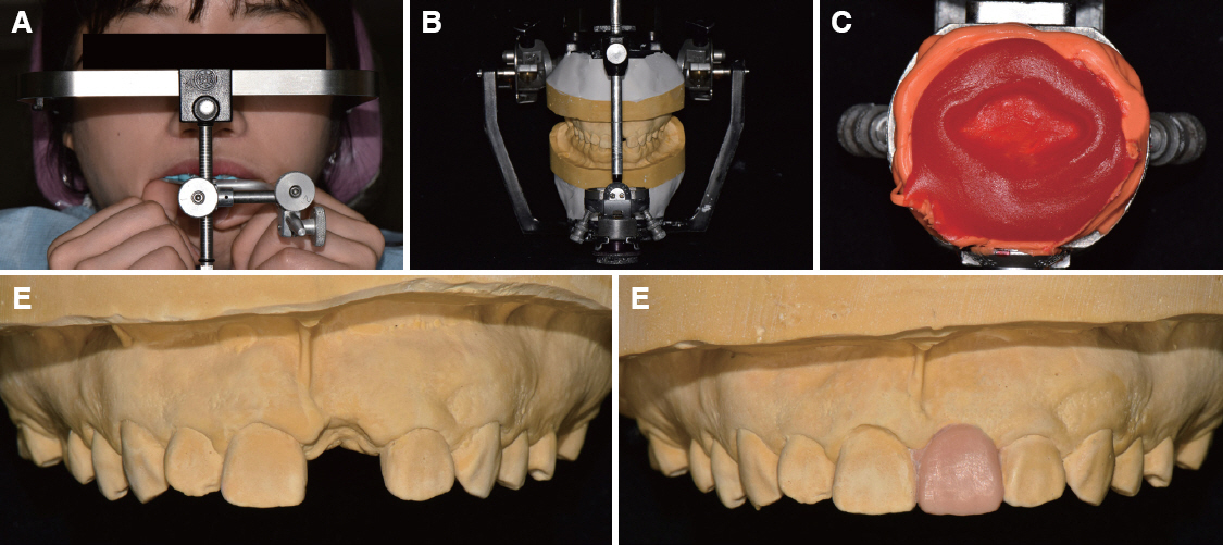
Fig. 2 Diagnostic wax-up model fabrication by using customized anterior guidance to maintain the occlusal pattern. (A) Face bow transfer, (B) Mounting of diagnostic model, (C) Customized anterior guidance table, (D) Diagnostic model of maxilla, (E) Diagnostic wax-up model of maxilla showing symmetric buccal surface.
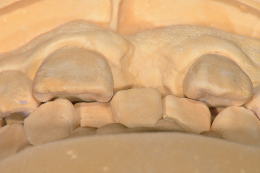
Fig. 3 Diagnositc model showing limited occlusal clearance due to crowding of mandibular anterior teeth.
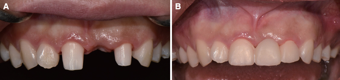
Fig. 4 Intraoral view after tooth preparation and temporary bridge restoration. (A) Tooth preparation, (B) Frontal view of temporary restoration.
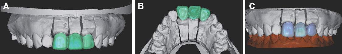
Fig. 5 Definitive prosthesis design by model scan and superimposition for duplicating the diagnostic wax-up. (A) Superimposed buccal image of definitive model and diagnostic wax-up model, (B) Superimposed lingual image of definitive model and diagnostic wax-up model, (C) Definitive prosthesis design.
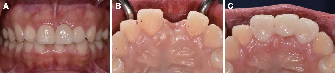
Fig. 6 Definitive prosthesis restoration and occlusion pattern by minimal adjustment. (A) Frontal view of definitive restoration, (B) Occlusion before treatment, (C) Maintenance of occlusion by duplicating the lingual surface of abutment teeth.
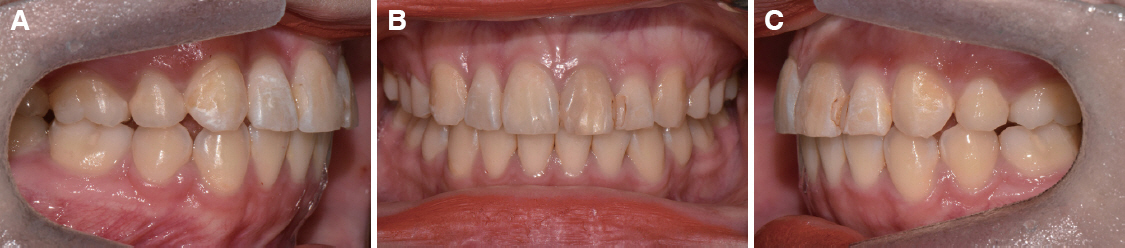
Fig. 7 Preoperative intraoral view showing the discoloration of #21. (A) Right lateral view, (B) Frontal view, (C) Left lateral view.
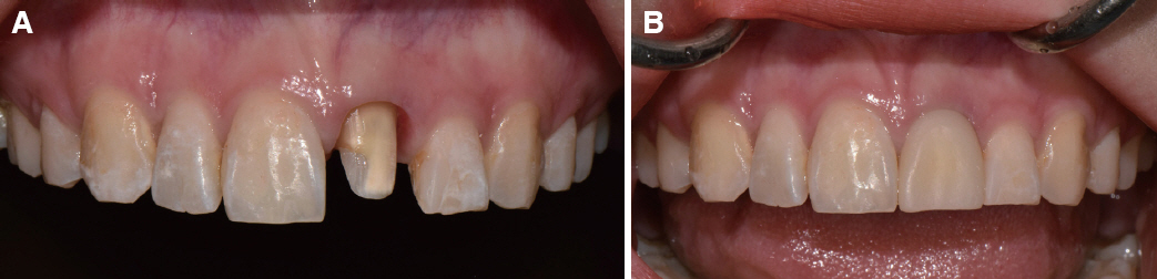
Fig. 8 Intraoral view after tooth preparation and temporary crown restoration. (A) Tooth preparation using putty index for uniform prosthesis thickness, (B) Frontal view of temporary crown restoration.

Fig. 9 Model scan and definitive prosthesis design by mirroring of #11 for symmetry. (A) Scan of definitive model, (B) Definitive prosthesis design showing esthetic appearance.
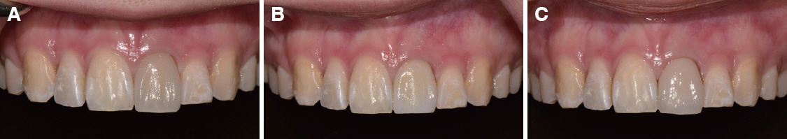
Fig. 10 Comparison of definitive prosthesis. (A) All-ceramic showing the failure of masking, (B) Monolithic zirconia showing the more esthetic appearance by masking the discoloration, (C) PFZ showing the failure of masking.
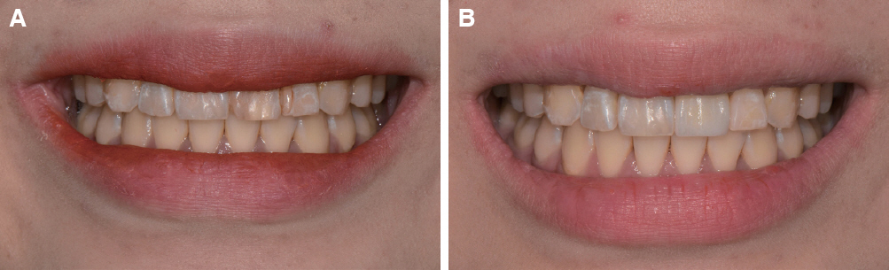
Fig. 11 Frontal view showing improved appearance during smile. (A) Before treatment, (B) After treatment.
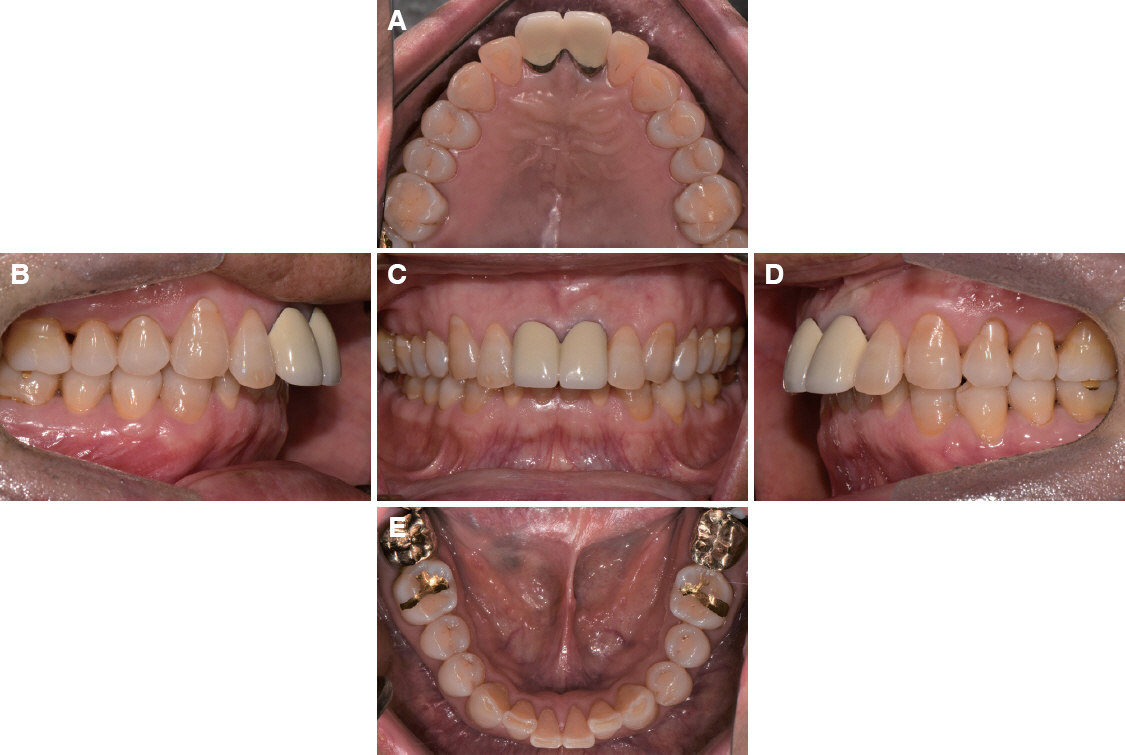
Fig. 12 Preoperative intraoral view showing the unesthetic prosthesis. (A) Occlusal view of maxilla, (B) Right lateral view, (C) Frontal view showing the unesthetic restoration and discoloration of gingiva, (D) Left lateral view, (E) Occlusal view of mandible.
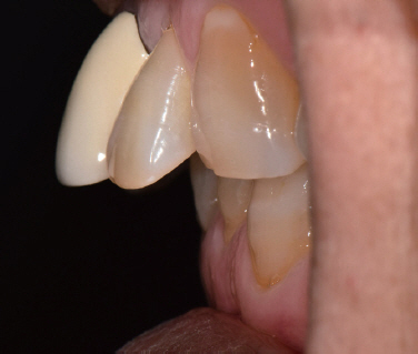
Fig. 13 Analysis of overjet. The overjet bigger than normal range.

Fig. 14 Fabrication of diagnostic wax-up model. (A) Before wax-up, (B) After wax-up reducing the overjet and modifying the axis of teeth.
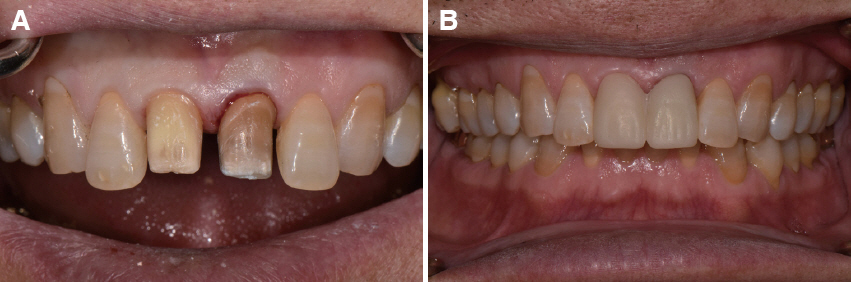
Fig. 15 Intraoral view after removal of prosthesis and temporary bridge restoration. (A) The discoloration of abutment teeth is shown after removal of prior prosthesis, (B) Frontal view of temporary bridge restoration.
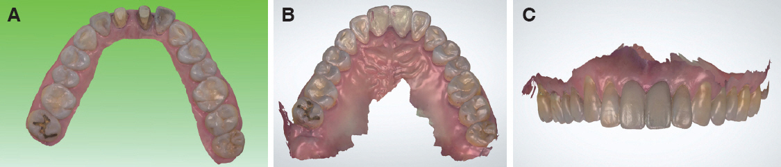
Fig. 16 Intraoral scan of teeth and temporary bridge for duplicating the temporary restoration. (A) Intraoral scan of teeth, (B) Lingual intraoral scan of temporary bridge, (C) Buccal intraoral scan of temporary bridge.

Fig. 17 Definitive prosthesis design by double scanning. (A) Superimposed buccal image of scan data, (B) Superimposed lingual image of scan data to maintain the occlusion pattern of temporary restoration.
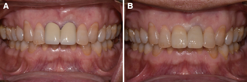
Fig. 18 Analysis of intraoral view. (A) Before treatment, (B) After treatment. Esthetics of prosthesis is improved and the discoloration of gingiva is relieved.
Reference
-
References
1. Bagby M, Marshall SJ, Marshall GW Jr. Metal ceramic compatibility: a review of the literature. J Prosthet Dent. 1990; 63:21–5. DOI: 10.1016/0022-3913(90)90259-F. PMID: 2404102.2. Isgrò G, Pallav P, van der Zel JM, Feilzer AJ. The influence of the veneering porcelain and different surface treatments on the biaxial flexural strength of a heat-pressed ceramic. J Prosthet Dent. 2003; 90:465–73. DOI: 10.1016/j.prosdent.2003.08.003. PMID: 14586311.3. Kim HK, Kim SH, Lee JB, Han JS, Yeo IS, Ha SR. Effect of the amount of thickness reduction on color and translucency of dental monolithic zirconia ceramics. J Adv Prosthodont. 2016; 8:37–42. DOI: 10.4047/jap.2016.8.1.37. PMID: 26949486. PMCID: PMC4769888.4. Teichmann M, Wienert AL, Rückbeil M, Weber V, Wolfart S, Edelhoff D. Ten-year survival and chipping rates and clinical quality grading of zirconiabased fixed dental prostheses. Clin Oral Investig. 2018; 22:2905–15. DOI: 10.1007/s00784-018-2378-1. PMID: 29520468.5. Stefanescu C, Ionita C, Nechita V, Drafta S, Oancea L, Petre A. Survival Rates and Complications for Zirconia-Based Fixed Dental Prostheses in a Period up to 10 Years: A Systematic Review. Eur J Prosthodont Restor Dent. 2018; 26:54–61. DOI: 10.1922/EJPRD_01681Stefanescu08. PMID: 29517875.6. Kwon SJ, Lawson NC, McLaren EE, Nejat AH, Burgess JO. Comparison of the mechanical properties of translucent zirconia and lithium disilicate. J Prosthet Dent. 2018; 120:132–7. DOI: 10.1016/j.prosdent.2017.08.004. PMID: 29310875.7. Tuncel İ, Turp I, Üşümez A. Evaluation of translucency of monolithic zirconia and framework zirconia materials. J Adv Prosthodont. 2016; 8:181–6. DOI: 10.4047/jap.2016.8.3.181. PMID: 27350851. PMCID: PMC4919487.8. Silva LHD, Lima E, Miranda RBP, Favero SS, Lohbauer U, Cesar PF. Dental ceramics: a review of new materials and processing methods. Braz Oral Res. 2017; 31:e58. DOI: 10.1590/1807-3107bor-2017.vol31.0058. PMID: 28902238.9. Zhang Y. Making yttria-stabilized tetragonal zirconia translucent. Dent Mater. 2014; 30:1195–203. DOI: 10.1016/j.dental.2014.08.375. PMID: 25193781.10. Huh YH, Yang EC, Park CJ, Cho LR. In vitro evaluation of the polishing effect and optical properties of monolithic zirconia. J Prosthet Dent. 2018; 119:994–9. DOI: 10.1016/j.prosdent.2017.06.015. PMID: 28965680.11. Tabatabaian F, Dalirani S, Namdari M. Effect of Thickness of Zirconia Ceramic on Its Masking Ability: An In Vitro Study. J Prosthodont. 2017; Apr. 28. doi:10.1111/jopr.12625. Epub ahead of print. DOI: 10.1111/jopr.12625. PMID: 28452411.12. Yang DH, Yang HS, Park SW, Lim HP, Yun KD, Vang MS. Full mouth implant rehabilitation with double scanning of provisional restoration. J Korean Acad Prosthodont. 2014; 52:252–7. DOI: 10.4047/jkap.2014.52.3.252.13. Hack GD, Sebastian B, Patzelt M. Evaluation of the accuracy of six intraoral scanning devices: An in-vitro investigation. ADA Professional Product Review. 2015; 10:1–5.14. Renne W, Ludlow M, Fryml J, Schurch Z, Mennito A, Kessler R, Lauer A. Evaluation of the accuracy of 7 digital scanners: An in vitro analysis based on 3-dimensional comparisons. J Prosthet Dent. 2017; 118:36–42. DOI: 10.1016/j.prosdent.2016.09.024. PMID: 28024822.
- Full Text Links
-
- Actions
-
Cited
- CITED
-
- Close
- Share
-
- Similar articles
-
- Full mouth rehabilitation using monolithic zirconia: a clinical report
- Full mouth rehabilitation with vertical increase in patient with severe tooth wear using monolithic zirconia prosthetic restoration
- Prosthetic treatment in esthetic area with monolithic zirconia using coloring liquid: a case report
- Restoration of anterior teeth with dental implant using multilayer zirconia
- Evaluation of translucency of monolithic zirconia and framework zirconia materials
