Chonnam Med J.
2016 Sep;52(3):219-219. 10.4068/cmj.2016.52.3.219.
A Case of Acute Pyelonephritis in Bilateral Renal Malrotation
- Affiliations
-
- 1Department of Internal Medicine, Chonnam National University Medical School, Gwangju, Korea. skimw@chonnam.ac.kr
- 2Department of Radiology, Chonnam National University Medical School, Gwangju, Korea.
- KMID: 2432274
- DOI: http://doi.org/10.4068/cmj.2016.52.3.219
Abstract
- No abstract available.
MeSH Terms
Figure
-
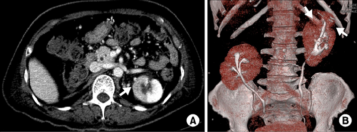
FIG. 1 An abdominal computed tomography axial image shows wedge-shaped low attenuation lesions in the left kidney at the upper pole, suggesting acute pyelonephritis (A). A three-dimensional computed tomography reconstruction image show anteriorly rotated bilateral kidneys (B).
Reference
-
1. Patil ST, Meshram MM, Kasote AP. Bilateral malrotation and lobulation of kidney with altered hilar anatomy: a rare congenital variation. Surg Radiol Anat. 2011; 33:941–944.
Article
- Full Text Links
-
- Actions
-
Cited
- CITED
-
- Close
- Share
-
- Similar articles
-
- A Rare Case of Acute Pyelonephritis Leading to Bilateral Subcapsular Renal Hematoma: A Case Report
- The Renal Scan in Acute Pyelonephritis
- A Case of Bilateral Xanthogranulomatous Pyelonephritis with Renal Failure
- A Case of Acute Renal Failure due to Bilateral Acute Pyelonephritis
- Bilateral Xanthogranulomatous Pyelonephritis in a Child
