Comparison of the effects of rapid maxillary expansion and alternate rapid maxillary expansion and constriction protocols followed by facemask therapy
- Affiliations
-
- 1Private Practice, Istanbul, Turkey. elvanonem89@gmail.com
- 2Department of Orthodontics, Faculty of Dentistry, Marmara University, Istanbul, Turkey.
- 3Department of Orthodontics, Faculty of Dentistry, Bezmialem Vakif University, Istanbul, Turkey.
- KMID: 2431080
- DOI: http://doi.org/10.4041/kjod.2019.49.1.49
Abstract
OBJECTIVE
The aim of this retrospective study was to evaluate and compare the changes in the pharyngeal airway (PA), maxillary sinus volume, and skeletal parameters after rapid maxillary expansion (RME) and alternate rapid maxillary expansion and constriction (Alt-RAMEC) followed by facemask (FM) therapy.
METHODS
The records of 40 patients with skeletal Class III malocclusion due to maxillary retrognathism were collected, and the patients were assigned into two groups. The first group comprised 8 male and 12 female patients (mean age, 10.0 ± 1.1 years) treated using RME/FM for an average of 10 months. The second group comprised 10 male and 10 female patients (mean age, 9.64 ± 1.3 years) treated using Alt-RAMEC/FM for an average of 12 months. Cone-beam computed tomography images acquired before (T0) and after treatment (T1) were evaluated.
RESULTS
Regarding the skeletal effects, significant differences between the groups were the increase in ANS-HRP (perpendicular distance of ANS to the horizontal reference plane, 0.99 mm, p <0.05) in the Alt-RAMEC/FM group and the decrease in PP-SN (palatal plane to Sella-Nasion plane, 0.93°, p < 0.05) in the RME/FM group. Maxillary sinus volumes increased significantly in both the groups, and the increase was statistically significantly higher in the Alt-RAMEC/FM group. Although no significant intergroup differences were observed in PA volumes, both lower (1,011.19 mm3) and total (1,601.21 mm3), PA volume increased significantly in the Alt-RAMEC/FM group.
CONCLUSIONS
The different expansion devices and protocols used with FM therapy do not seem to affect the forward movement of the maxilla and PA volumes. In contrast, the increase in maxillary sinus volume was greater in the Alt-RAMEC/FM protocol.
Keyword
MeSH Terms
Figure
-

Figure 1 The flow diagram of patient selection based on the CONSORT statement guidelines. FM, Facemask; RME, rapid maxillary expansion; Alt-RAMEC, alternate rapid maxillary expansion and constriction; 3D, three-dimensional.
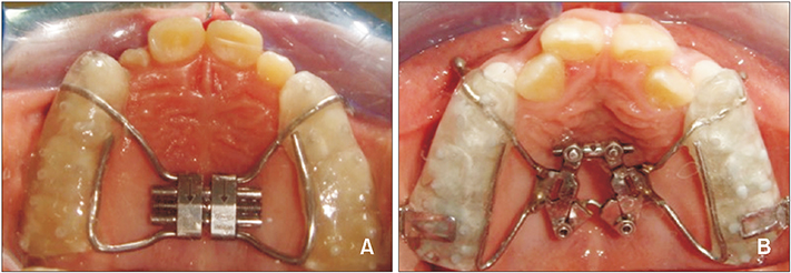
Figure 2 A, Intraoral photo of the Hyrax screw. B, Intraoral photo of the double-hinged expansion screw.

Figure 3 Skeletal measurements. 1, PP-SN (°); 2, GoMe-SN (°); 3, SNA (°); 4, SNB (°); 5, ANB (°); 6, FMA (°); 7, AVRP (mm); 8, B-VRP (mm); 9, ANS-HRP (mm); 10, PNSHRP (mm); 11, S-Go (mm); 12, ANS-Me (mm); 13, N-Me (mm). PP-SN, The angle between palatal plane and sella-nasion plane; GoMe-SN, the angle between gonion-menton plane and sella-nasion plane; SNA, the angle between sella, nasion and A points; SNB, the angle between sella, nasion and B points; ANB, the angle between A, nasion and B points; FMA, the angle between Frankfort horizontal reference plane and mandibular plane; A-VRP, the distance between A point and the vertical reference plane; B-VRP, the distance between B point and the vertical reference plane; ANS-HRP, the distance between anterior nasal spine point and the horizontal reference plane; PNS-HRP, the distance between posterior nasal spine point and the horizontal reference plane; S-Go, the distance between sella and gonion points; ANS-Me, the distance between anterior nasal spine and menton points; N-Me, the distance between nasion and menton points.
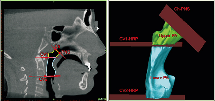
Figure 4 Three-dimensional reconstruction of the pharyngeal airway (PA). Ch, The point before the last slice where the posterior wall of the pharynx and the nasal septum fuse; CV1, the most anterior and inferior points of the first cervical vertebra; CV2, the most anterior and inferior points of the second cervical vertebra; PNS, posterior nasal spine; HRP, horizontal reference plane.
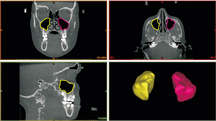
Figure 5 Three-dimensional reconstruction of the maxillary sinuses.
Cited by 2 articles
-
Does surgically assisted maxillary protraction with skeletal anchorage and Class III elastics affect the pharyngeal airway? A retrospective, long-term study
Elvan Onem Ozbilen, Petros Papaefthymiou, Hanife Nuray Yilmaz, Nazan Küçükkeleş
Korean J Orthod. 2023;53(1):35-44. doi: 10.4041/kjod22.117.Comparison of changes in the nasal cavity, pharyngeal airway, and maxillary sinus volumes after expansion and maxillary protraction with two protocols: Rapid palatal expansion versus alternate rapid maxillary expansion and constriction
Weitao Liu, Shaonan Zhou, Edwin Yen, Bingshuang Zou
Korean J Orthod. 2023;53(3):175-184. doi: 10.4041/kjod22.075.
Reference
-
1. Keles A, Tokmak EC, Erverdi N, Nanda R. Effect of varying the force direction on maxillary orthopedic protraction. Angle Orthod. 2002; 72:387–396.2. Ellis E 3rd, McNamara JA Jr. Components of adult Class III malocclusion. J Oral Maxillofac Surg. 1984; 42:295–305.
Article3. Kama JD, Ozer T, Baran S. Orthodontic and orthopaedic changes associated with treatment in subjects with Class III malocclusions. Eur J Orthod. 2006; 28:496–502.
Article4. Liou EJ, Tsai WC. A new protocol for maxillary protraction in cleft patients: repetitive weekly protocol of alternate rapid maxillary expansions and constrictions. Cleft Palate Craniofac J. 2005; 42:121–127.
Article5. Kaya D, Kocadereli I, Kan B, Tasar F. Effects of facemask treatment anchored with miniplates after alternate rapid maxillary expansions and constrictions; a pilot study. Angle Orthod. 2011; 81:639–646.
Article6. Oktay H, Ulukaya E. Maxillary protraction appliance effect on the size of the upper airway passage. Angle Orthod. 2008; 78:209–214.
Article7. Kaygisiz E, Tuncer BB, Yüksel S, Tuncer C, Yildiz C. Effects of maxillary protraction and fixed appliance therapy on the pharyngeal airway. Angle Orthod. 2009; 79:660–667.
Article8. Kilinç AS, Arslan SG, Kama JD, Ozer T, Dari O. Effects on the sagittal pharyngeal dimensions of protraction and rapid palatal expansion in Class III malocclusion subjects. Eur J Orthod. 2008; 30:61–66.
Article9. Sayinsu K, Isik F, Arun T. Sagittal airway dimensions following maxillary protraction: a pilot study. Eur J Orthod. 2006; 28:184–189.
Article10. Hiyama S, Suda N, Ishii-Suzuki M, Tsuiki S, Ogawa M, Suzuki S, et al. Effects of maxillary protraction on craniofacial structures and upper-airway dimension. Angle Orthod. 2002; 72:43–47.11. Mucedero M, Baccetti T, Franchi L, Cozza P. Effects of maxillary protraction with or without expansion on the sagittal pharyngeal dimensions in Class III subjects. Am J Orthod Dentofacial Orthop. 2009; 135:777–781.
Article12. Pamporakis P, Nevzatoğlu Ş, Küçükkeleş N. Three-dimensional alterations in pharyngeal airway and maxillary sinus volumes in Class III maxillary deficiency subjects undergoing orthopedic facemask treatment. Angle Orthod. 2014; 84:701–707.
Article13. Celikoglu M, Buyukcavus MH. Changes in pharyngeal airway dimensions and hyoid bone position after maxillary protraction with different alternate rapid maxillary expansion and construction protocols: a prospective clinical study. Angle Orthod. 2017; 87:519–525.
Article14. Aboudara C, Nielsen I, Huang JC, Maki K, Miller AJ, Hatcher D. Comparison of airway space with conventional lateral headfilms and 3-dimensional reconstruction from cone-beam computed tomography. Am J Orthod Dentofacial Orthop. 2009; 135:468–479.
Article15. Yilmaz BS, Kucukkeles N. Skeletal, soft tissue, and airway changes following the alternate maxillary expansions and constrictions protocol. Angle Orthod. 2014; 84:868–877.
Article16. Gautam P, Valiathan A, Adhikari R. Skeletal response to maxillary protraction with and without maxillary expansion: a finite element study. Am J Orthod Dentofacial Orthop. 2009; 135:723–728.
Article17. Canturk BH, Celikoglu M. Comparison of the effects of face mask treatment started simultaneously and after the completion of the alternate rapid maxillary expansion and constriction procedure. Angle Orthod. 2015; 85:284–291.
Article18. The SEDENTEXCT Project. Radiation protection: cone beam CT for dental and maxillofacial radiology: evidence based guidelines 2011 (v2.0 final) [Internet]. The SEDENTEXCT Project;2011. Available from: www.sedentexct.eu/files/guidelines_final.pdf.19. El H, Palomo JM. Airway volume for different dentofacial skeletal patterns. Am J Orthod Dentofacial Orthop. 2011; 139:e511–e521.
Article20. Iwasaki T, Hayasaki H, Takemoto Y, Kanomi R, Yamasaki Y. Oropharyngeal airway in children with Class III malocclusion evaluated by cone-beam computed tomography. Am J Orthod Dentofacial Orthop. 2009; 136:318.e1–318.e9. discussion 318-9.
Article21. Ngan P, Hägg U, Yiu C, Merwin D, Wei SH. Soft tissue and dentoskeletal profile changes associated with maxillary expansion and protraction headgear treatment. Am J Orthod Dentofacial Orthop. 1996; 109:38–49.
Article22. Baik HS. Clinical results of the maxillary protraction in Korean children. Am J Orthod Dentofacial Orthop. 1995; 108:583–592.
Article23. Kapust AJ, Sinclair PM, Turley PK. Cephalometric effects of face mask/expansion therapy in Class III children: a comparison of three age groups. Am J Orthod Dentofacial Orthop. 1998; 113:204–212.
Article24. Cordasco G, Matarese G, Rustico L, Fastuca S, Caprioglio A, Lindauer SJ, et al. Efficacy of orthopedic treatment with protraction facemask on skeletal Class III malocclusion: a systematic review and meta-analysis. Orthod Craniofac Res. 2014; 17:133–143.
Article25. Yüksel S, Uçem TT, Keykubat A. Early and late facemask therapy. Eur J Orthod. 2001; 23:559–568.
Article26. Li H, Lu X, Shi J, Shi H. Measurements of normal upper airway assessed by 3-dimensional computed tomography in Chinese children and adolescents. Int J Pediatr Otorhinolaryngol. 2011; 75:1240–1246.
Article27. Chen X, Liu D, Liu J, Wu Z, Xie Y, Li L, et al. Three-dimensional evaluation of the upper airway morphological changes in growing patients with skeletal Class III malocclusion treated by protraction headgear and rapid palatal expansion: a comparative research. PLoS One. 2015; 10:e0135273.
Article28. Taylor M, Hans MG, Strohl KP, Nelson S, Broadbent BH. Soft tissue growth of the oropharynx. Angle Orthod. 1996; 66:393–400.29. Barghouth G, Prior JO, Lepori D, Duvoisin B, Schnyder P, Gudinchet F. Paranasal sinuses in children: size evaluation of maxillary, sphenoid, and frontal sinuses by magnetic resonance imaging and proposal of volume index percentile curves. Eur Radiol. 2002; 12:1451–1458.
Article30. Park IH, Song JS, Choi H, Kim TH, Hoon S, Lee SH, et al. Volumetric study in the development of paranasal sinuses by CT imaging in Asian: a pilot study. Int J Pediatr Otorhinolaryngol. 2010; 74:1347–1350.
Article
- Full Text Links
-
- Actions
-
Cited
- CITED
-
- Close
- Share
-
- Similar articles
-
- Comparison of changes in the nasal cavity, pharyngeal airway, and maxillary sinus volumes after expansion and maxillary protraction with two protocols: Rapid palatal expansion versus alternate rapid maxillary expansion and constriction
- A posteroanterior cephalometric study on the change of maxilla by rapid palatal expansion
- A study on the effect of rapid maxillary expansion and its relapse
- Salvage rapid maxillary expansion for the relapse of maxillary transverse expansion after Le Fort I with parasagittal osteotomy
- Crowding with no posterior crossbite treatment byrapid palatal expansion
