Ann Dermatol.
2019 Feb;31(1):106-107. 10.5021/ad.2019.31.1.106.
A Case of Ochronosis with Atypical Manifestations Involving the Perioral Area and Sclera
- Affiliations
-
- 1Department of Dermatology, Chonnam National University Medical School, Gwangju, Korea. jbmlee@jnu.ac.kr
- KMID: 2430815
- DOI: http://doi.org/10.5021/ad.2019.31.1.106
Abstract
- No abstract available.
MeSH Terms
Figure
-
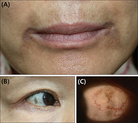
Fig. 1 Asymptomatic dark brownish ill-defined patches around the perioral area (A), and hyperpigmentation of both sclerae (B: right eye, C: left eye).
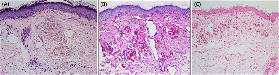
Fig. 2 (A) Histopathological examination shows amorphous brownish banana-shaped fibers in the dermis. No specific findings in the epidermis (H&E, ×200). (B) Immunohistochemical staining reveals negative results with Periodic acid-Schiff (×200), and (C) ferric acid stain negative (×40).
Reference
-
1. Kramer KE, Lopez A, Stefanato CM, Phillips TJ. Exogenous ochronosis. J Am Acad Dermatol. 2000; 42:869–871.
Article2. Khaled A, Kerkeni N, Hawilo A, Fazaa B, Kamoun MR. Endogenous ochronosis: case report and a systematic review of the literature. Int J Dermatol. 2011; 50:262–267.
Article3. Levin CY, Maibach H. Exogenous ochronosis. An update on clinical features, causative agents and treatment options. Am J Clin Dermatol. 2001; 2:213–217.4. Skorin L Jr, Turpin S. Minocycline-induced hyperpigmentation of the skin, sclera, and palpebral conjunctiva. Can J Ophthalmol. 2017; 52:e79–e81.
Article
- Full Text Links
-
- Actions
-
Cited
- CITED
-
- Close
- Share
-
- Similar articles
-
- A Case of Perioral Dermatitis Involving the Eyelids
- Atypical Cheiro-oral Syndrome Presented With Bilateral Perioral Sensory Symptoms in Unilateral Thalamic Infarction
- A Case of Childhood Granulomatous Perioral Dermatitis
- Ochronotic Arthropathy: Degenerative and Complex Tear of Black Meniscus
- A Case of Granulomatous Perioral Dermatitis
