Yonsei Med J.
2005 Dec;46(6):794-798.
A Bile Based Study of Clonorchis sinensis Infections in Patients with Biliary Tract Diseases in Ulsan, Korea
- Affiliations
-
- 1Department of Internal Medicine, Kyung Hee University College of Medicine, Seoul, Korea.
- 2Department of Internal Medicine, Ulsan University Hospital, University of Ulsan College of Medicine, Ulsan, Korea.
Abstract
- Stool examination is believed to be the most reliable method for detecting Clonorchis sinensis (CS) eggs. However, it has limited value for diagnosing clonorchiasis when the biliary tract is obstructed or when there is a light infection. We evaluated the infection states of CS in patients with biliary tract diseases using a bile sample. From January 2001 to August 2003, 238 patients who had undergone endoscopic biliary drainage were prospectively included in the study. The patients' bile samples were obtained directly from the nasobiliary drainage tube and then analyzed to detect CS eggs. The overall CS egg positive rate was 28.2% (35.4% in males, 19.4% in females). The egg positive rate was similar in all age groups examined: 26.7% in 30-39 years, 25.0% in 40-49 years, 24.4% in 50-59 years, 30.2% in 60-69 years, 35.3% in 70-79 years, and 25.0% in 80 years of age and over. There were no significant differences in the egg positive rate between the disease groups: 32.6% in bile duct cancer, 38.5% in gallbladder cancer, and 26.4% in gallstone diseases. Our results show that the CS infection rate was very high, regardless of the age, gender, and type of diseases of the patients. Although the study population was limited to patients with biliary tract diseases, it is assumed that clonorchiasis is still an endemic disease in Ulsan, Korea.
Keyword
MeSH Terms
Figure
-
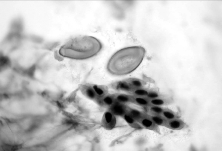
Fig. 1 Eggs of the liver fluke Clonorchis sinensis in the bile specimen. The prominent operculum that produces the "shoulders" characteristic of eggs of this species is evident (Papanicolaou's stain, ×1000).
Reference
-
1. Rim HJ. Clonorchiasis in Korea. Korean J Parasitol. 1990. 28:63–78.2. Choi MH, Ryu JS, Lee M, Li S, Chung BS, Chai JY, et al. Specific and common antigens of Clonorchis sinensis and Opisthorchis viverrini (Opisthorchidae, Trematoda). Korean J Parasitol. 2003. 41:155–163.3. Joo KR, Lee MS, Yun TG, Choo YI, Lee JW, Jo YC, et al. Diagnostic usefulness of intradermal test for clonorchiasis in patients with pancreatobiliary diseases. Korean J Gastroenterol. 2003. 42:232–236.4. Joo CY. Epidemiological studies of Clonorchis sinensis in vicinity of river Taewha, Kyungnam province, Korea. Korean J Parasitol. 1980. 18:199–214.5. Wang KX, Zhang RB, Cui YB, Tian Y, Cai R, Li CP. Clinical and epidemiological data of patients with clonorchiasis. World J Gastroenterol. 2004. 10:446–448.6. Yu SH, Kawanaka M, Li XM, Xu LQ, Lan CG, Rui L. Epidemiological investigation on Clonorchis sinensis in human population in an area of South China. Jpn J Infect Dis. 2003. 56:168–171.7. Kim BJ, Yeon JW, Ock MS. Infection rates of Enterobius vermicularis and Clonorchis sinensis of primary school children in Hamyang-gun, Gyeongsangnam-do (province), Korea. Korean J Parasitol. 2001. 39:323–325.8. Carpenter HA. Bacterial and parasitic choloangitis. Mayo Clin Proc. 1998. 73:473–478.9. Liu LX, Harinasuta KT. Liver and intestinal flukes. Gastroenterol Clin North Am. 1996. 25:627–636.10. Kim BJ, Ock MS, Kim IS, Yeo UB. Infection status of Clonorchis sinensis in residents of Hamyang-gun, Gyeongsangnam-do, Korea. Korean J Parasitol. 2002. 40:191–193.11. Lee GS, Cho IS, Lee YH, Noh HJ, Shin DW, Lee SG, et al. Epidemiological study of clonorchiasis and metagonimiasis along the Geum-gang (River) in Okcheon-gun (county), Korea. Korean J Parasitol. 2002. 40:9–16.12. Ministry of Health and Welfare and Korea Association of Health. The Sixth Report. Prevalence of intestinal parasitic infections in Korea. 1997. Seoul, Korea:13. Shin BM, Choi KI. Diagnostic significance of intradermal test compared with radiologic findings for clonorchiasis. Korean J Clin Pathol. 2000. 20:81–86.14. Lin YL, Chen ER, Yen CM. Antibodies in serum of patients with clonorchiasis before and after treatment. Southeast Asian J Trop Med Public Health. 1995. 26:114–119.15. Hong ST, Park KH, Seo M, Choi BI, Chai JY, Lee SH. Correlation of sonograhic findings with histopathological changes of the bile ducts in rabbits infected with Clonorchis sinensis. Korean J Parasitol. 1994. 32:223–230.16. Lim JH. Radiologic findings of clonorchiasis. AJR Am J Roentgenol. 1990. 155:1001–1008.17. Choi TK, Wong KP, Wong J. Cholangiographic appearance in clonorchiasis. Br J Radiol. 1984. 57:681–684.
- Full Text Links
-
- Actions
-
Cited
- CITED
-
- Close
- Share
-
- Similar articles
-
- A Case of Clonorchis Sinensis Infection in the Gastric Pylorus Associated with Gastric Adenocarcinoma
- Infestation state of clonorchis sinensis in patients with pancreatobiliary diseases in ulsan: based on bile examination
- Two cases of Nephrotic Syndrome Associated with Kimura's Disease
- Computed tomography in evaluation of jaundiced patients due to clonorchis sinensis
- ERCP Findings In Hepatic Clonorchiasis
