J Korean Soc Radiol.
2018 Dec;79(6):340-347. 10.3348/jksr.2018.79.6.340.
Value of Image Subtraction for the Identification of Hepatocellular Carcinoma Capsule on Gadoxetic Acid-Enhanced MRI
- Affiliations
-
- 1Department of Radiology, Yonsei University Wonju College of Medicine, Wonju Severance Christian Hospital, Wonju, Korea. radajh@yonsei.ac.kr
- 2Center of Biomedical Data Science, Yonsei University Wonju College of Medicine, Wonju Severance Christian Hospital, Wonju, Korea.
- KMID: 2427383
- DOI: http://doi.org/10.3348/jksr.2018.79.6.340
Abstract
- PURPOSE
To evaluate value of image subtraction for identifying hepatocellular carcinoma (HCC) capsule on gadoxetic acid-enhanced MR images.
MATERIALS AND METHODS
This study involved 108 patients at risk of HCC preoperatively examined using gadoxetic acid-enhanced MRI with hepatic resection between May 2015 and February 2017. We evaluated qualities of subtraction images and presence of capsular appearance on portal venous or transitional phases conventional and subtraction images. We assessed effect of capsular appearance on subtraction images on HCC.
RESULTS
After excluding 1 patient who had treated by transarterial chemoembolization prior to surgery and 33 patients with unsatisfactory subtraction image qualities, 82 focal hepatic lesions (73 HCC, 5 non-HCC malignancies, and 4 benign) from 74 patients were analyzed. Regarding detection of capsules, sensitivity, accuracy, and area under the receiver operating characteristic curve (AUC) on subtraction images were significantly higher than those on conventional images (95.4%, 89.0%, and 0.80, respectively; p < 0.001), though specificities were same (64.7%). For diagnosis of HCC, sensitivity, accuracy, and AUC on subtraction images were significantly higher than on conventional images (82.2%, 79.3%, and 0.69, respectively; p = 0.011), though specificities were identical (55.6%).
CONCLUSION
Portal venous or transitional phase gadoxetic acid-enhanced MRI subtraction images could improve detection of HCC capsule.
MeSH Terms
Figure
-
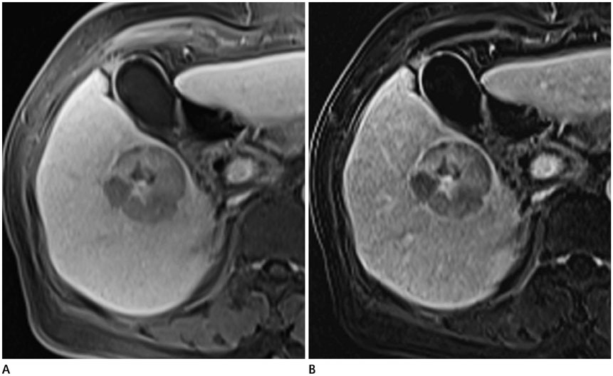
Fig. 1 Case of a 73-year-old woman with hepatocellular carcinoma encased by a histopathologically confirmed fibrous capsule in the right liver A. A conventional capsular appearance is not well visualized in the conventional TP image. B. However, a smooth hyperintense rim is readily detected on the subtracted TP image. TP = transitional phase
Reference
-
1. El-Serag HB, Davila JA, Petersen NJ, McGlynn KA. The continuing increase in the incidence of hepatocellular carcinoma in the United States: an update. Ann Intern Med. 2003; 139:817–823.
Article2. Kojiro M. Histopathology of liver cancers. Best Pract Res Clin Gastroenterol. 2005; 19:39–62.
Article3. Kadoya M, Matsui O, Takashima T, Nonomura A. Hepatocellular carcinoma: correlation of MR imaging and histopathologic findings. Radiology. 1992; 183:819–825.
Article4. Ishigami K, Yoshimitsu K, Nishihara Y, Irie H, Asayama Y, Tajima T, et al. Hepatocellular carcinoma with a pseudocapsule on gadolinium-enhanced MR images: correlation with histopathologic findings. Radiology. 2009; 250:435–443.
Article5. Organ Procurement and Transplantation Network. OPTN/UNOS policy 9.3.G.iv. Accessed Mar 16, 2016. Available at: http://optn.transplant.hrsa.gov/ContentDocuments/OPTN_Policies.pdf-nameddest=Policy_09. Published Jan 1, 2015.6. Liver Reporting and Data System, version 2013.1. American College of Radiology;Accessed Mar 16, 2016. Available at: http://www.acr.org/Quality-Safety/Resources/LIRADS/.7. Miraglia R, Pietrosi G, Maruzzelli L, Petridis I, Caruso S, Marrone G, et al. Predictive factors of tumor response to trans-catheter treatment in cirrhotic patients with hepatocellular carcinoma: a multivariate analysis of pre-treatment findings. World J Gastroenterol. 2007; 13:6022–6026.
Article8. Ng IO, Lai EC, Ng MM, Fan ST. Tumor encapsulation in hepatocellular carcinoma. A pathologic study of 189 cases. Cancer. 1992; 70:45–49.
Article9. Grazioli L, Olivetti L, Fugazzola C, Benetti A, Stanga C, Dettori E, et al. The pseudocapsule in hepatocellular carcinoma: correlation between dynamic MR imaging and pathology. Eur Radiol. 1999; 9:62–67.
Article10. Choi JY, Lee JM, Sirlin CB. CT and MR imaging diagnosis and staging of hepatocellular carcinoma: part II. Extracellular agents, hepatobiliary agents, and ancillary imaging features. Radiology. 2014; 273:30–50.
Article11. Dioguardi Burgio M, Picone D, Cabibbo G, Midiri M, Lagalla R, Brancatelli G. MR-imaging features of hepatocellular carcinoma capsule appearance in cirrhotic liver: comparison of gadoxetic acid and gadobenate dimeglumine. Abdom Radiol (NY). 2016; 41:1546–1554.
Article12. Hope TA, Fowler KJ, Sirlin CB, Costa EA, Yee J, Yeh BM, et al. Hepatobiliary agents and their role in LI-RADS. Abdom Imaging. 2015; 40:613–625.
Article13. Yu JS, Kim YH, Rofsky NM. Dynamic subtraction magnetic resonance imaging of cirrhotic liver: assessment of high signal intensity lesions on nonenhanced T1-weighted images. J Comput Assist Tomogr. 2005; 29:51–58.14. Yu JS, Rofsky NM. Dynamic subtraction MR imaging of the liver: advantages and pitfalls. AJR Am J Roentgenol. 2003; 180:1351–1357.
Article15. Seçil M, Obuz F, Altay C, Gencel O, Iğci E, Sağol O, et al. The role of dynamic subtraction MRI in detection of hepatocellular carcinoma. Diagn Interv Radiol. 2008; 14:200–204.16. Sundarakumar DK, Wilson GJ, Osman SF, Zaidi SF, Maki JH. Evaluation of image registration in subtracted 3D dynamic contrast-enhanced MRI of treated hepatocellular carcinoma. AJR Am J Roentgenol. 2015; 204:287–296.
Article17. Cho ES, Choi JY. MRI features of hepatocellular carcinoma related to biologic behavior. Korean J Radiol. 2015; 16:449–464.
Article18. Asayama Y, Nishie A, Ishigami K, Ushijima Y, Takayama Y, Fujita N, et al. Distinguishing intrahepatic cholangiocarcinoma from poorly differentiated hepatocellular carcinoma using precontrast and gadoxetic acid-enhanced MRI. Diagn Interv Radiol. 2015; 21:96–104.
Article19. Khan AS, Hussain HK, Johnson TD, Weadock WJ, Pelletier SJ, Marrero JA. Value of delayed hypointensity and delayed enhancing rim in magnetic resonance imaging diagnosis of small hepatocellular carcinoma in the cirrhotic liver. J Magn Reson Imaging. 2010; 32:360–366.
Article20. Park HJ, Jang KM, Kang TW, Song KD, Kim SH, Kim YK, et al. Identification of imaging predictors discriminating different primary liver tumours in patients with chronic liver disease on gadoxetic acid-enhanced MRI: a classification tree analysis. Eur Radiol. 2016; 26:3102–3111.
Article21. Rimola J, Forner A, Tremosini S, Reig M, Vilana R, Bianchi L, et al. Non-invasive diagnosis of hepatocellular carcinoma ≤ 2 cm in cirrhosis. Diagnostic accuracy assessing fat, capsule and signal intensity at dynamic MRI. J Hepatol. 2012; 56:1317–1323.22. Suh YJ, Kim MJ, Choi JY, Park YN, Park MS, Kim KW. Differentiation of hepatic hyperintense lesions seen on gadoxetic acid-enhanced hepatobiliary phase MRI. AJR Am J Roentgenol. 2011; 197:W44–W52.
Article23. Vogl TJ, Kümmel S, Hammerstingl R, Schellenbeck M, Schumacher G, Balzer T, et al. Liver tumors: comparison of MR imaging with Gd-EOB-DTPA and Gd-DTPA. Radiology. 1996; 200:59–67.
Article24. An C, Rhee H, Han K, Choi JY, Park YN, Park MS, et al. Added value of smooth hypointense rim in the hepatobiliary phase of gadoxetic acid-enhanced MRI in identifying tumour capsule and diagnosing hepatocellular carcinoma. Eur Radiol. 2017; 27:2610–2618.
Article
- Full Text Links
-
- Actions
-
Cited
- CITED
-
- Close
- Share
-
- Similar articles
-
- Diagnosis of Hepatocellular Carcinoma with Gadoxetic Acid-Enhanced MRI: 2016 Consensus Recommendations of the Korean Society of Abdominal Radiology
- Gadoxetic acid-enhanced magnetic resonance imaging: Hepatocellular carcinoma and mimickers
- Gadoxetic Acid (Gd-EOB-DTPA)-Enhanced MRI versus Gadobenate Dimeglumine (Gd-BOPTA)-Enhanced MRI for Preoperatively Detecting Hepatocellular Carcinoma: an Initial Experience
- Hepatic Angiomyolipoma Presenting as a Hyperintense Lesion During the Hepatobiliary Phase of Gadoxetic Acid Enhanced-MRI: a Case Report
- Usefulness of Arterial Subtraction in Applying Liver Imaging Reporting and Data System (LI-RADS) Treatment Response Algorithm to Gadoxetic Acid-Enhanced MRI
