Value and Clinical Application of Orthopedic Metal Artifact Reduction Algorithm in CT Scans after Orthopedic Metal Implantation
- Affiliations
-
- 1Department of Radiology, Shengjing Hospital of China Medical University, Shenyang, Liaoning Province 110004, China. guoqy@sj-hospital.org
- 2Department of Orthopedics, Shengjing Hospital of China Medical University, Shenyang, Liaoning Province 110004, China.
- KMID: 2427308
- DOI: http://doi.org/10.3348/kjr.2017.18.3.526
Abstract
OBJECTIVE
To evaluate orthopedic metal artifact reduction algorithm (O-MAR) in CT orthopedic metal artifact reduction at different tube voltages, identify an appropriate low tube voltage for clinical practice, and investigate its clinical application.
MATERIALS AND METHODS
The institutional ethical committee approved all the animal procedures. A stainless-steel plate and four screws were implanted into the femurs of three Japanese white rabbits. Preoperative CT was performed at 120 kVp without O-MAR reconstruction, and postoperative CT was performed at 80-140 kVp with O-MAR. Muscular CT attenuation, artifact index (AI) and signal-to-noise ratio (SNR) were compared between preoperative and postoperative images (unpaired t test), between paired O-MAR and non-O-MAR images (paired Student t test) and among different kVp settings (repeated measures ANOVA). Artifacts' severity, muscular homogeneity, visibility of inter-muscular space and definition of bony structures were subjectively evaluated and compared (Wilcoxon rank-sum test). In the clinical study, 20 patients undertook CT scan at low kVp with O-MAR with informed consent. The diagnostic satisfaction of clinical images was subjectively assessed.
RESULTS
Animal experiments showed that the use of O-MAR resulted in accurate CT attenuation, lower AI, better SNR, and higher subjective scores (p < 0.010) at all tube voltages. O-MAR images at 100 kVp had almost the same AI and SNR as non-O-MAR images at 140 kVp. All O-MAR images were scored ≥ 3. In addition, 95% of clinical CT images performed at 100 kVp were considered satisfactory.
CONCLUSION
O-MAR can effectively reduce orthopedic metal artifacts at different tube voltages, and facilitates low-tube-voltage CT for patients with orthopedic metal implants.
MeSH Terms
Figure
-

Fig. 1 CT imaging of rabbit femur by thick multiple planar reconstruction to show relationship between femur and metal implants.Plate was placed parallel to femur, and axis of screw was perpendicular to femur, as much as possible.
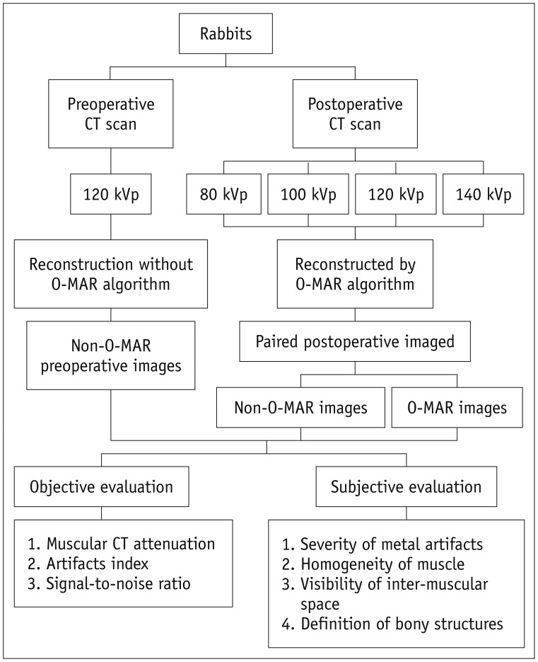
Fig. 2 Schema of CT performance and image evaluation in animal experiments.O-MAR = orthopedic metal artifact reduction algorithm
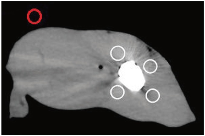
Fig. 3 Schematic of ROIs selection.Four white circles showed ROIs drawn in muscular area around metal implants. Red circle was used for measurements in background region away from metal implants. ROIs = regions of interest
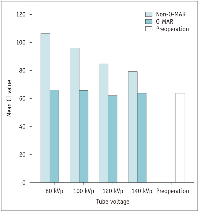
Fig. 4 Comparison of muscular CT attenuation (n = 24).O-MAR = orthopedic metal artifact reduction
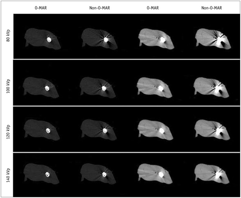
Fig. 5 Subjective evaluation of image quality (n = 24).O-MAR images were better than non-O-MAR images under all tube voltages. Image quality was better with increased tube voltage on both O-MAR and non-O-MAR images. O-MAR = orthopedic metal artifact reduction
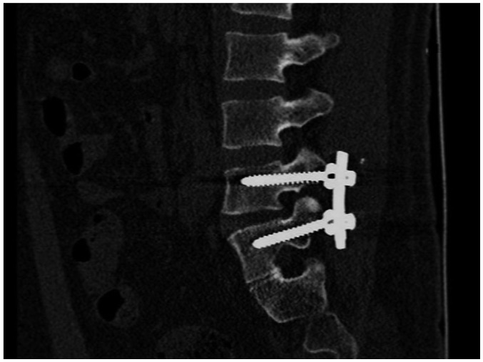
Fig. 6 Metal implants in lumbar spine.Little artifact without obvious distortion and clear cancellous bone were shown. It was subjectively scored 4 points for both severity of metal artifacts and definition of bony structures.
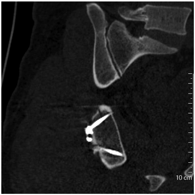
Fig. 7 Metal implants in pelvis.Little artifact without obvious distortion and clear cancellous bone were shown. This image was subjectively scored 4 points. Little osteoporosis was found surrounding metal, and fracture line was still clear.
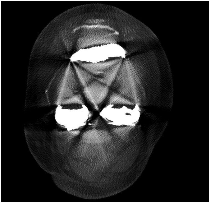
Fig. 8 Metal implants in knee.Severe artifacts with obvious distortion were shown, resulting in blurry appearance of both bony structures and metal implants. It was subjectively scored 1 point for severity of metal artifacts and definition of bony structures.
Cited by 1 articles
-
Contrast-Enhanced CT with Knowledge-Based Iterative Model Reconstruction for the Evaluation of Parotid Gland Tumors: A Feasibility Study
Chae Jung Park, Ki Wook Kim, Ho-Joon Lee, Myeong-Jin Kim, Jinna Kim
Korean J Radiol. 2018;19(5):957-964. doi: 10.3348/kjr.2018.19.5.957.
Reference
-
1. Buckwalter KA, Rydberg J, Kopecky KK, Crow K, Yang EL. Musculoskeletal imaging with multislice CT. AJR Am J Roentgenol. 2001; 176:979–986. PMID: 11264094.
Article2. White LM, Buckwalter KA. Technical considerations: CT and MR imaging in the postoperative orthopedic patient. Semin Musculoskelet Radiol. 2002; 6:5–17. PMID: 11917267.
Article3. Kim M, Choi YS, Kim H, Choi NH. Postoperative evaluation after anterior cruciate ligament reconstruction: measurements and abnormalities on radiographic and CT imaging. Korean J Radiol. 2016; 17:919–930. PMID: 27833408.
Article4. Rinkel J, Dillon WP, Funk T, Gould R, Prevrhal S. Computed tomographic metal artifact reduction for the detection and quantitation of small features near large metallic implants: a comparison of published methods. J Comput Assist Tomogr. 2008; 32:621–629. PMID: 18664852.5. Abdoli M, Ay MR, Ahmadian A, Zaidi H. A virtual sinogram method to reduce dental metallic implant artefacts in computed tomography-based attenuation correction for PET. Nucl Med Commun. 2010; 31:22–31. PMID: 19829166.
Article6. Andersen AH, Kak AC. Simultaneous algebraic reconstruction technique (SART): a superior implementation of the art algorithm. Ultrason Imaging. 1984; 6:81–94. PMID: 6548059.
Article7. Gilbert P. Iterative methods for the three-dimensional reconstruction of an object from projections. J Theor Biol. 1972; 36:105–117. PMID: 5070894.
Article8. Metal Artifact Reduction for Orthopedic Implants (O-MAR). updated Jan 8, 2012. Accessed January 14, 2016. Available at: http://clinical.netforum.healthcare.philips.com/us_en/Explore/White-Papers/CT/Metal-Artifact-Reduction-for-Orthopedic-Implants-(O-MAR)#.9. Kidoh M, Nakaura T, Nakamura S, Tokuyasu S, Osakabe H, Harada K, et al. Reduction of dental metallic artefacts in CT: value of a newly developed algorithm for metal artefact reduction (O-MAR). Clin Radiol. 2014; 69:e11–e16. PMID: 24156796.
Article10. Jeong S, Kim SH, Hwang EJ, Shin CI, Han JK, Choi BI. Usefulness of a metal artifact reduction algorithm for orthopedic implants in abdominal CT: phantom and clinical study results. AJR Am J Roentgenol. 2015; 204:307–317. PMID: 25615752.
Article11. Li H, Noel C, Chen H, Harold Li H, Low D, Moore K, et al. Clinical evaluation of a commercial orthopedic metal artifact reduction tool for CT simulations in radiation therapy. Med Phys. 2012; 39:7507–7517. PMID: 23231300.
Article12. Barrett JF, Keat N. Artifacts in CT: recognition and avoidance. Radiographics. 2004; 24:1679–1691. PMID: 15537976.
Article13. Lee MJ, Kim S, Lee SA, Song HT, Huh YM, Kim DH, et al. Overcoming artifacts from metallic orthopedic implants at high-field-strength MR imaging and multi-detector CT. Radiographics. 2007; 27:791–803. PMID: 17495293.
Article14. Pessis E, Campagna R, Sverzut JM, Bach F, Rodallec M, Guerini H, et al. Virtual monochromatic spectral imaging with fast kilovoltage switching: reduction of metal artifacts at CT. Radiographics. 2013; 33:573–583. PMID: 23479714.
Article15. Li B, Yadava G, Hsieh J. Quantification of head and body CTDI(VOL) of dual-energy x-ray CT with fast-kVp switching. Med Phys. 2011; 38:2595–2601. PMID: 21776796.16. Brook OR, Gourtsoyianni S, Brook A, Mahadevan A, Wilcox C, Raptopoulos V. Spectral CT with metal artifacts reduction software for improvement of tumor visibility in the vicinity of gold fiducial markers. Radiology. 2012; 263:696–705. PMID: 22416251.
Article17. Müller J, Buzug TM. Spurious structures created by interpolation-based CT metal artifact reduction. Spie Med Imaging. 2009; 7258:72581Y.
Article18. De Man B, Nuyts J, Dupont P, Marchal G, Suetens P. Metal streak artifacts in X-ray computed tomography: a simulation study. IEEE Trans Nucl Sci. 1999; 46:691–696.
Article19. Meyer E, Raupach R, Lell M, Schmidt B, Kachelriess M. Normalized metal artifact reduction (NMAR) in computed tomography. Med Phys. 2010; 37:5482–5493. PMID: 21089784.
Article20. Wang G, Frei T, Vannier MW. Fast iterative algorithm for metal artifact reduction in X-ray CT. Acad Radiol. 2000; 7:607–614. PMID: 10952111.
Article21. Wang G, Snyder DL, O'Sullivan JA, Vannier MW. Iterative deblurring for CT metal artifact reduction. IEEE Trans Med Imaging. 1996; 15:657–664. PMID: 18215947.22. De Man B. Iterative reconstruction for reduction of metal artifacts in computed tomography [dissertation]. Leuven: Katholieke Universiteit Leuven;2001.23. Huang JY, Kerns JR, Nute JL, Liu X, Balter PA, Stingo FC, et al. An evaluation of three commercially available metal artifact reduction methods for CT imaging. Phys Med Biol. 2015; 60:1047–1067. PMID: 25585685.
Article
- Full Text Links
-
- Actions
-
Cited
- CITED
-
- Close
- Share
-
- Similar articles
-
- Fluorodeoxyglucose Positron-Emission Tomography/Computed Tomography and Magnetic Resonance Imaging for Adverse Local Tissue Reactions near Metal Implants after Total Hip Arthroplasty: A Preliminary Report
- Reduction of Metal Artifact around Titanium Alloy-based Pedicle Screws on CT Scan Images: An Approach using a Digital Image Enhancement Technique
- Effect of object position in the field of view and application of a metal artifact reduction algorithm on the detection of vertical root fractures on cone-beam computed tomography scans: An in vitro study
- Comparison of the Quality of Various Polychromatic and Monochromatic Dual-Energy CT Images with or without a Metal Artifact Reduction Algorithm to Evaluate Total Knee Arthroplasty
- Comparison of Metal Artifact Reduction for Orthopedic Implants versus Standard Filtered Back Projection: Value of Postoperative CT after Hip Replacement
