Anat Cell Biol.
2015 Sep;48(3):201-204. 10.5115/acb.2015.48.3.201.
Rectus and oblique muscles of eyeball: a morphometric study of Indian population
- Affiliations
-
- 1Department of Anatomy, All India Institute of Medical Sciences, Bhopal, Madhya Pradesh, India. sunita.anatomy@aiimsbhopal.edu.in
- KMID: 2424823
- DOI: http://doi.org/10.5115/acb.2015.48.3.201
Abstract
- During the strengthening and weakening procedures of intraocular muscles, distance of insertion from the sclerocorneal junction is an important determinant in the identification of muscles. During repositioning of the aponeurosis of the muscles, it is desired that the width should not change in order to avoid diversion of forces. Available anatomic studies on insertions of extraocular muscles are few, date back to early twentieth century and have been conducted on mostly white population. The present study is an attempt to document the insertions of recti and oblique muscles in Indian population. Forty eyeballs were removed from orbit. Insertion of recti and obliqui were cleaned and eyeballs were perfused with normal saline to regain the volume (hence shape and size) before recording observations. Insertion of recti and obliqui muscles were observed under various study parameters. The distance of insertion of recti from the limbus were found to be 7.3 mm, 8.06 mm, 8.71 mm, and 8.74 mm for medial, inferior, lateral, and superior rectus, respectively. The superior oblique was aponeurotic and found to be more variable in mode of insertion as compared to inferior oblique which had a fleshy and relatively constant insertion. The observations on insertion of recti and obliqui as obtained in present study differ from earlier studies to the tune of 1-1.5 mm. This may be attributed to adoption of method of reperfusion of eyeball before recording observations thus maintaining size close to in vivo. The observations are expected to be closer to actual.
Keyword
MeSH Terms
Figure
-
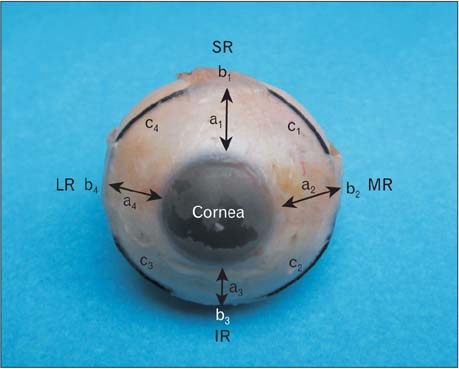
Fig. 1 Showing insertions of rectus muscles and various study parameters. a1, a2, a3, and a4 represent distance of insertion from limbus of SR, MR, IR, and LR, respectively. b1, b2, b3, and b4 represents width of insertions of SR, MR, IR, and LR, respectively. c1, c2, c3, and c4 represent distance between adjacent recti, respectively. MR, medial rectus; IR, inferior rectus; LR, lateral rectus; SR, superior rectus.
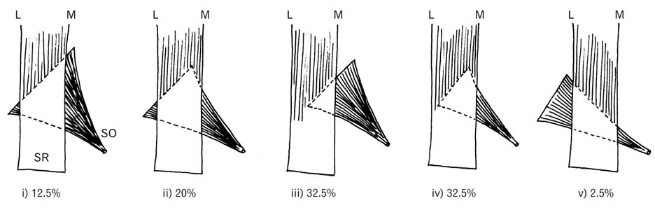
Fig. 2 Shows different pattern of insertion of SO with respect to SR. SO, superior oblique; SR, superior rectus; L, lateral; M, medial.
Reference
-
1. Standring S, Ellis H, Healy JC, Johnson D, Williams A, Collins P, Wigley C. Gray's anatomy: the anatomical basis of clinical practice. 39th ed. London: Churchill Livingstone;2005. p. 91–694.2. Last RJ. Anatomy, regional and applied. 7th ed. Edinburgh: Churchill Livingstone;1984. p. 432–440.3. Kanski JJ. Clinical ophthalmology: a systematic approach. 6th ed. London: Elsevier Butterworth-Heinemann;2007. p. 780–782.4. Roper-Hall G, Cruz OA, Chung SM. Results of extraocular muscle surgery in WEBINO bilateral internuclear ophthalmoplegia patients. J AAPOS. 2008; 12:277–281.5. Apt L. An anatomical reevaluation of rectus muscle insertions. Trans Am Ophthalmol Soc. 1980; 78:365–375.6. Sevel D. The origins and insertions of the extraocular muscles: development, histologic features, and clinical significance. Trans Am Ophthalmol Soc. 1986; 84:488–526.7. Khurana AK. Comprehensive ophthalmology. 3rd ed. New Delhi: New Age International Publishers;2003. p. 127.8. Jaggi GP, Laeng HR, Müntener M, Killer HE. The anatomy of the muscle insertion (scleromuscular junction) of the lateral and medial rectus muscle in humans. Invest Ophthalmol Vis Sci. 2005; 46:2258–2263.
- Full Text Links
-
- Actions
-
Cited
- CITED
-
- Close
- Share
-
- Similar articles
-
- The Quantitative Measurement of the Oculocardiac Reflex
- Anatomical Evaluation of Extraocular Muscles and Relationships Among Macula and Optic Nerve in Enucleated Eye
- A Case of Simultaneous Injury to Ipsilateral Superior Rectus and Superior Oblique Muscle
- Surgical Correction of Exotropia due to Oculomotor Nerve Palsy
- Strabismus Surgery on Congenital Oculomotor Nerve Palsied Eye
