Long-term Changes in the Peripapillary RNFL and Macular GCIPL Thicknesses after Panretinal Photocoagulation in Diabetic Retinopathy Patients
- Affiliations
-
- 1Department of Ophthalmology, School of Medicine, Kyungpook National University, Daegu, Korea. proector97@gmail.com
- 2Department of Ophthalmology, Kyungpook National University Hospital, Daegu, Korea.
- KMID: 2422582
- DOI: http://doi.org/10.3341/jkos.2018.59.10.938
Abstract
- PURPOSE
To investigate longitudinal changes in the thicknesses of the peripapillary retinal nerve fiber layer (pRNFL) and the macular ganglion cell-inner plexiform layer (mGCIPL) in patients with diabetic retinopathy 3 years after panretinal photocoagulation (PRP).
METHODS
We retrospectively reviewed the medical records of 60 eyes of 35 patients who were diagnosed with diabetic retinopathy and treated with PRP. The pRNFL and mGCIPL thicknesses were measured by optical coherence tomography at baseline, and then at 1, 3, 6, 9, 12, 24, and 36 months after PRP.
RESULTS
The pRNFL and mGCIPL thicknesses (average and all sections) at 1 year after PRP increased significantly from baseline (p < 0.05, respectively). The average pRNFL and mGCIPL thicknesses showed a tendency to decrease continuously from 2 years after PRP (p < 0.05, respectively). There was no statistically significant difference in the average thicknesses of the pRNFL and the mGCIPL between pre-PRP (92.27 ± 7.76 µm, and 85.00 ± 4.80 µm, respectively) and 3 years after PRP (93.93 ± 7.49 µm, and 81.87 ± 14.00 µm, respectively) (p = 0.121, and p = 0.622, respectively).
CONCLUSIONS
Although the pRNFL and the mGCIPL thicknesses increased at 1 year after PRP, there was no statistical difference in the average thicknesses of the pRNFL and the mGCIPL between pre-PRP and 3 years after PRP. These results should be considered with respect to the diagnosis and progression of glaucoma in patients with diabetic retinopathy who undergo PRP.
Keyword
MeSH Terms
Figure
-

Figure 1 Peripapillary retinal nerve fiber layer (RNFL) and macular ganglion cell-inner plexiform layer (GCIPL) thickness in Cirrus optical coherence tomography (OCT). (A) OCT shows the RNFL thicknesses of average and four-quadrant in both eyes. (B) OCT shows GCIPL thicknesses of average and six-sector in both eyes. OD = oculus dexter; OS = oculus sinister; GCL = ganglion cell layer; IPL = inner plexiform layer; S = superior; T = temporal; N = nasal; I = inferior; NA = non-available.

Figure 2 Longitudinal changes in the average and four-quadrant thicknesses (µm) of the peripapillary retinal nerve fiber layer (RNFL) after panretinal photocoagulation (PRP). The RNFL thicknesses (average and four-quadrant) increased at 1 month after PRP and had declined continuously since then (A–E). Repeated-measures analysis of variance corrected by Bonferroni method showed that the RNFL thicknessess (average and four-quadrant) increased at 1 year after PRP (all p < 0.05), but there was no statistically significant difference of the average, inferior, nasal and temporal thicknesses of the RNFL between pre-PRP and 3 years after PRP (A, C–E). The superior RNFL thickness (93.93 ± 7.49 µm) decreased at 3 years after PRP from the thickness (92.27 ± 7.76 µm) before PRP (p = 0.442) (B). *p < 0.05 by repeated-measures analysis of variance corrected by the Bonferroni methods; †p < 0.05 by paired t-test.
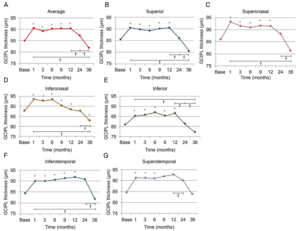
Figure 3 Longitudinal changes in the average and six-sector thicknesses (µm) of the macular ganglion cell-inner plexiform layer (GCIPL) after panretinal photocoagulation (PRP). Repeated-measures analysis of variance corrected by Bonferroni method showed that macular GCIPL thicknesses (average and 6-sector) increased in 1 year after PRP (all p < 0.05). The average GCIPL thickness showed a tendency to increase until 1 year, but to decrease continuously from 2 year after PRP. There was no statistically significant difference of the average, superior, superonasal, inferior, inferotemporal and superotemporal GCIPL thicknesses between pre-PRP and 3 years after PRP (A–C, E–G). The inferonasal GCIPL thickness (81.87 ± 19.0 µm) decreased at 3 years after PRP from the thickness (85.00 ± 4.80 µm) before PRP (p = 0.544) (D). *p < 0.05 by repeated-measures analysis of variance corrected by the Bonferroni methods; †p < 0.05 by by paired t-test.
Reference
-
1. Barber AJ. A new view of diabetic retinopathy: a neurodegenerative disease of the eye. Prog Neuropsychopharmacol Biol Psychiatry. 2003; 27:283–290.
Article2. Stefanson E. Oxygen and diabetic eye disease. Graefes Arch Clin Exp Ophthalmol. 1990; 228:120–123.3. Frisch GD, Shawaluk PD, Adams DO. Remote nerve fibre bundle alterations in the retina as caused by argon laser photocoagulation. Nature. 1974; 248:433–435.
Article4. Retineour RJ, Kozousek V, Chauhan BC. The effect of panretinal photocoagulation for diabetic retinopathy on retinal nerve fiber layer thickness and optic disc topography. Br J Ophthalmol. 2009; 93:838–839.5. Kim J, Woo SJ, Ahn J, et al. Long-term temporal changes of peripapillary retinal nerve fiber layer thickness before and after panretinal photocoagulation in severe diabetic retinopathy. Retina. 2012; 32:2052–2060.
Article6. Mwanza JC, Budenz DL, Godfrey DG, et al. Diagnostic performance of optical coherence tomography ganglion cell--inner plexiform layer thickness measurements in early glaucoma. J Ophthalmology. 2014; 121:849–854.
Article7. Sihota R, Sony P, Gupta V, et al. Diagnostic capability of optical coherence tomography in evaluating the degree of glaumcomatous retinal nerve fiber damage. Invest Ophthalmo Vis Sci. 2006; 47:2006–2010.8. Mwanza JC, Durbin MK, Budenz DL, et al. Profile and predictors of normal ganglion cell-inner plexiform layer thickness measured with frequency-domain optical coherence tomography. Invest Ophthalmol Vis Sci. 2011; 52:7872–7879.
Article9. Mwanza JC, Oakley JD, Budenz DL, et al. Macular ganglion cell-inner plexiform layer: automated detection and thickness reproducibility with spectral domain-optical coherence tomography in glaucoma. Invest Ophthalmol Vis Sci ophthalmol. 2011; 52:8323–8329.
Article10. Kim JJ, Im JC, Shin JP, et al. One-year follow-up of macular ganglion cell layer and peripapillary retinal nerve fiber layer thickness changes after panretinal photocoagulation. Br J ophthalmol. 2014; 98:213–217.11. Roohipoor R, Dantism S, Ahmadrji A, et al. Subfoveal choroidal thickness after panretinal photocoagulation with red and green lase in bilateral proliferative diabetic retinopathy patients: short term results. J Ophthalmol. 2016; 2016:9364861.12. Park YR, Jee D. Changes in peripapillary retinal nerve fiber layer thickness after pattern scanning laser photocoagulation in patients with diabetic retinopathy. Korean J Ophthalmol. 2014; 28:220–225.
Article13. Shin JS, Lee YH. Changes in macular retinal layers and peripapillary nerve fiber layer thickness after 577-nm pattern scanning laser in patients with diabetic retinopathy. Korean J Ophthalmol. 2017; 31:497–507.
Article14. Mendívil A, Cuartero V, Mendívil MP. Ocular blood flow velocities in patients with proliferative diabetic retinopathy before and after scatter photocoagulation: a prospective study. Eur J Ophthalmol. 1995; 5:259–264.
Article15. Hiroshiba N, Ogura Y, Nishiwaki H, et al. Alterations of retinal microcirculation in response to scatter photocoagulation. Invest Ophthalmol Vis Sci. 1998; 39:769–776.16. Nonaka A, Kiryu J, Tsujikawa A, et al. Inflammatory response after scatter laser photocoagulated retina. Invest Ophthalmol Vis Sci. 2002; 43:1204–1209.17. Itaya M, Sakurai E, Nozaki M, et al. Upregulation of VEGF in murine retina via monocyte recruitment after retinal scatter laser photocoagulation. Invest Ophthalmol Vis Sci. 2007; 48:5677–5683.
Article18. Jampol LM, Odia I, Glassman AR, et al. Panretinal photocoagulation versus ranibizumab for proliferative diabetic retinopathy comparison of peripapillary retinal nerve fiber layer thickness in a randomized clinical trial. Retina. 2017; DOI: 10.1097/IAE.0000000000001909.19. Gillies MC, Su T, Stayt J, et al. Effect of high glucose on permeability of retinal capillary endothelium in vitro. Invest Ophthalmol Vis Sci. 1997; 38:635–642.20. Rungger-Brändle E, Dosso AA, Leuenberger PM. Glial reactivity, an early feature of diabetic retinopathy. Invest Ophthalmol Vis Sci. 2000; 41:1971–1980.21. Kim JT, Lee JK, Moon NJ, Cho HK. Analysis of the optic nerve head and RNFL thickness using optical coherence tomography in diabetes. J Korean Ophthalmol Soc. 2008; 49:935–941.
Article22. Apple DJ, Wyhinny GJ, Goldenberg MF, et al. Experimental argon laser photocoagulation. I. Effects on retinal nerve fiber layer. Arch Ophthalmol. 1976; 94:137–144.23. Yang HS, Kim JG, Cha JB, et al. Quantitative analysis of neural tissues around the optic disc after panretinal photocoagulation in patients with diabetic retinopathy. Plos one. 2017; 12:e0186229. eCollection 2017.
Article24. Tso MO, Wallow IH, Elgin S. Experimental photocoagulation of the human retina: I. Correlation of physical, clinical, and pathologic data. Arch Ophthalmol. 1977; 95:1035–1040.25. Van Dijk HW, Verbraak FD, Stehouwer M, et al. Association of visual function and ganglion cell layer thickness in patients with diabetes mellitus type 1 and no or minimal diabetic retinopathy. Vision Res. 2011; 51:224–228.
Article26. Lee SB, Kwag JY, Lee HJ, et al. The longitudinal changes of retinal nerve fiber layer thickness after panretinal photocoagulation in diabetic retinopathy patients. Retina. 2013; 33:188–193.
Article27. Maeshima K, Utsugi-Sutah N, Otani T, Kishi S. Progressive enlargement of scattered photocoagulation scars in diabetic retinopathy. Retina. 2004; 24:507–511.
Article28. Shin YJ, Kyoung HS, Park KH, Yu HG. The analysis of retinal nerve fiber layer in the patients with nonproliferative diabetic retinopathy. J Korean Ophthalmol Soc. 2003; 44:2010–2015.
- Full Text Links
-
- Actions
-
Cited
- CITED
-
- Close
- Share
-
- Similar articles
-
- Macular Thickness and Visual Acuity Before and After Panretinal Photocoagulation in Severe Diabetic Retinopathy
- Short-term Effect of Intravitreal Bevacizumab Injection Preventing Panretinal Photocoagulation-Induced Macular Edema in Proliferative Diabetic Retinopathy
- The Effects of Pa n retinal Photocoagulation on Macular Microcirculation in Diabetic Retinopathy(Short term follow up)
- Changes in Macular Retinal Layers and Peripapillary Nerve Fiber Layer Thickness after 577-nm Pattern Scanning Laser in Patients with Diabetic Retinopathy
- Peripapillary Retinal Nerve Fiber Layer Thickness Change After Panretinal Photocoagulation in Patients With Diabetic Retinopathy
