Anterior Choroidal Artery Aneurysms: Influence of Regional Microsurgical Anatomy on Safety of Endovascular Treatment
- Affiliations
-
- 1Department of Neurosurgery, Baylor College of Medicine, Houston, TX, USA. Peter.Kan@bcm.edu
- KMID: 2422560
- DOI: http://doi.org/10.7461/jcen.2018.20.1.47
Abstract
- Several anatomical variables critically influence therapeutic strategizing for anterior choroidal artery (AChA) aneurysms, and specifically, the safety of flow diversion for these lesions. We review the microsurgical anatomy of the AChA, discussing and detailing these considerations in the treatment of AChA aneurysms, theoretically and in the light of our recent findings.
Figure
-
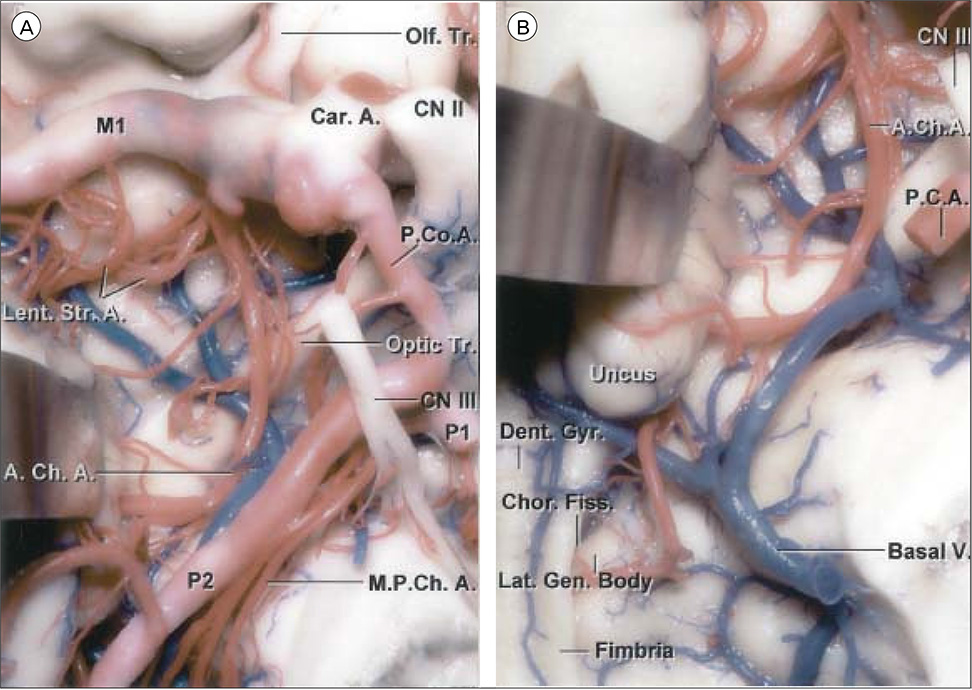
Fig. 1 Anterior choroidal artery course and relations viewed from inferiorly. (A) The right AChA arises from the posterior wall of the ICA above the origin of the PCoA and passes backward below the optic tract and lateral to the PCA. It ascends around the medial surface of the uncus as it travels posteriorly. (B) The posterior uncal segment has been retracted. The AChA passes above the posterior uncal segment and enters the temporal horn by passing through the choroidal fissure located between the thalamus above and fimbria of the fornix below. The lateral geniculate body forms the part of the thalamus above where the artery enters the choroidal fissure. The dentate gyrus is located at the lower edge of the fimbria. Reprinted with permission from reference 17. Olf. Tr. = FULL NAME; M1 = sphenoidal segment of middle cerebral artery; Car. A. = internal carotid artery; CN II = optic nerve; Lent. Str. A. = lenticulostriate arteries; P.Co.A = posterior communicating artery; Optic Tr. = optic tract; CN III = oculomotor nerve; P1 = pre-communicating segment of posterior cerebral artery; A.Ch.A = anterior choroidal artery; P2 = post-communicating segment of posterior cerebral artery; M.P.Ch.A = medial posterior choroidal artery; P.C.A. = posterior cerebral artery; Dent. Gyr. = dentate gyrus; Chor. Fiss. = choroidal fissure; Lat. Gen. Body = lateral geniculate body; Basal V = basal vein of Rosenthal; ICA = internal carotid artery.
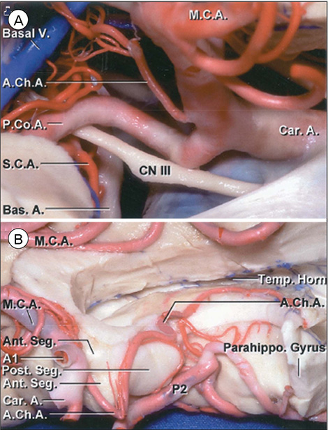
Fig. 2 Anterior choroidal artery course and relations viewed from laterally and medially. (A) Lateral view. The right AChA arises above the origin of the PComA and passes upward and backward around the uncus to reach the temporal horn. (B) Medial view. The AChA pursues an angulated course, descending along the anterior segment of the uncus, but at the uncal apex, it turns sharply upward, reaching the upper part of the posterior uncal segment before entering the temporal horn. Reprinted with permission from reference 17. Basal V. = basal vein of Rosenthal; M.C.A. = middle cerebral artery; A.Ch.A. = anterior choroidal artery; P.Co.A = posterior communicating artery; Car. A. = internal carotid artery; S.C.A. = FULL NAME; CN III = oculomotor nerve; cating segment of anterior cerebral artery; Bas. A. = basilar artery; Temp. Horn = temporal horn of lateral ventricle; Ant. Seg. = anterior segment of uncus; A1 = pre-communicating segment of anterior cerebral artery; Parahippo. Gyrus = parahippocampal gyrus; Post. Seg. = posterior segment of uncus; P2 = second segment of posterior cerebral artery; PComA = FULL NAME.
Reference
-
1. Abbie AA. The clinical significance of the anterior choroidal artery. Brain. 1933; 09. 56:233–246.
Article2. Carpenter MB, Noback CR, Moss ML. The anterior choroidal artery; its origins course, distribution, and variations. AMA Arch Neurol Psychiatry. 1954; 06. 71(6):714–722.3. Cooper IS. Surgical alleviation of Parkinsonism; effects of occlusion of the anterior choroidal artery. J Am Geriatr Soc. 1954; 11. 2(11):691–718.
Article4. Cooper IS. Surgical occlusion of the anterior choroidal artery in Parkinsonism. Surg Gynecol Obstet. 1954; 08. 99(2):207–219.5. Decroix JP, Graveleau P, Masson M, Cambier J. Infarction in the territory of the anterior choroidal artery. A clinical and computerized tomographic study of 16 cases. Brain. 1986; 12. 109(Pt 6):1071–1085.6. Erdem A, Yaşargil G, Roth P. Microsurgical anatomy of the hippocampal arteries. J Neurosurg. 1993; 08. 79(2):256–265.
Article7. Fernández-Miranda JC, de Oliveira E, Rubino PA, Wen HT, Rhoton AL Jr. Microvascular anatomy of the medial temporal region: part 1: its application to arteriovenous malformation surgery. Neurosurgery. 2010; 09. 67:3 Suppl Operative. ons237–ons276. discussion ons276.
Article8. Gibo H, Carver CC, Rhoton AL Jr, Lenkey C, Mitchell RJ. Microsurgical anatomy of the middle cerebral artery. J Neurosurg. 1981; 02. 54(2):151–169.
Article9. Gibo H, Lenkey C, Rhoton AL Jr. Microsurgical anatomy of the supraclinoid portion of the internal carotid artery. J Neurosurg. 1981; 10. 55(4):560–574.
Article10. Huther G, Dörfl J, Van der Loos H, Jeanmonod D. Microanatomic and vascular aspects of the temporomesial region. Neurosurgery. 1998; 11. 43(5):1118–1136.
Article11. Inci S, Arat A, Ozgen T. Distal anterior choroidal artery aneurysms. Surg Neurol. 2007; 01. 67(1):46–52. discussion 52.
Article12. Marinković S, Gibo H, Brigante L, Nikodijević I, Petrović P. The surgical anatomy of the perforating branches of the anterior choroidal artery. Surg Neurol. 1999; 07. 52(1):30–36.
Article13. Morandi X, Brassier G, Darnault P, Mercier P, Scarabin JM, Duval JM. Microsurgical anatomy of the anterior choroidal artery. Surg Radiol Anat. 1996; 18(4):275–280.
Article14. Nelles M, Gieseke J, Flacke S, Lachenmayer L, Schild HH, Urbach H. Diffusion tensor pyramidal tractography in patients with anterior choroidal artery infarcts. AJNR Am J Neuroradiol. 2008; 03. 29(3):488–493.
Article15. Perlmutter D, Rhoton AL Jr. Microsurgical anatomy of the anterior cerebral-anterior communicating-recurrent artery complex. J Neurosurg. 1976; 09. 45(3):259–272.
Article16. Rand RW, Brown WJ, Stern WE. Surgical occlusion of the anterior choroidal arteries in Parkinsonism; clinical and neuropathologic findings. Neurology. 1956; 06. 6(6):390–401.17. Rhoton AL Jr. The supratentorial arteries. Neurosurgery. 2002; 10. 51:4 Suppl. S53–S120.
Article18. Rhoton AL Jr, Fujii K, Fradd B. Microsurgical anatomy of the anterior choroidal artery. Surg Neurol. 1979; 08. 12(2):171–187.19. Ribas EC, Yagmurlu K, Wen HT, Rhoton AL Jr. Microsurgical anatomy of the inferior limiting insular sulcus and the temporal stem. J Neurosurg. 2015; 06. 122(6):1263–1273.
Article20. Rosner SS, Rhoton AL Jr, Ono M, Barry M. Microsurgical anatomy of the anterior perforating arteries. J Neurosurg. 1984; 09. 61(3):468–485.
Article21. Saeki N, Rhoton AL Jr. Microsurgical anatomy of the upper basilar artery and the posterior circle of Willis. J Neurosurg. 1977; 05. 46(5):563–578.
Article22. Srinivasan VM, Ghali MGZ, Cherian J, Mokin M, Puri AS, Grandhi R, et al. Flow diversion for anterior choroidal artery (AChA) aneurysms: a multi-institutional experience. J Neurointerv Surg. 2017; 10. 31. [Epub ahead of print].
Article23. Tanriover N, Kucukyuruk B, Ulu MO, Isler C, Sam B, Abuzayed B, et al. Microsurgical anatomy of the cisternal anterior choroidal artery with special emphasis on the preoptic and postoptic subdivisions. J Neurosurg. 2014; 05. 120(5):1217–1228.
Article24. van der Zwan A, Hillen B, Tulleken CA, Dujovny M, Dragovic L. Variability of the territories of the major cerebral arteries. J Neurosurg. 1992; 12. 77(6):927–940.
Article25. Yaşargil MG. Anterior choroidal artery. In : Yaşargil MG, editor. Microsurgical Anatomy of the Basal Cisterns and Vessels of the Brain, Diagnostic Studies, General Operative Techniques and Pathological Considerations of the Intracranial Aneurysms. Stuttgart: Georg Thieme Verlag;1984. p. 66–70.
- Full Text Links
-
- Actions
-
Cited
- CITED
-
- Close
- Share
-
- Similar articles
-
- Microsurgical anatomy of the Anterior Cerebral-anterior Communicating Artery
- Microsurgical Anatomy of the Basilar Artery: Surgical Approaches to the Basilar Trunk and Vertebrobasilar Junction Aneurysms
- Clinical Experiences of Anterior Choroidal Artery Aneurysm
- Flow recovery after posterior clinoidectomy for surgical clipping of anterior choroidal aneurysm
- Classification of Anterior Communicating Artery Aneurysm with Regard to its Microsurgical Anatomy
