Intrahepatic Pseudoaneurysms Complicating Transjugular Liver Biopsy in Liver Transplantation Patients: Three Case Reports
- Affiliations
-
- 1Department of Radiology and Research Institute of Radiology, Asan Medical Center, University of Ulsan College of Medicine, Seoul, Korea. kogy@amc.seoul.kr
Abstract
- An intrahepatic pseudoaneurysm is a rare complication following transjugular liver biopsy. Transarterial embolization is considered a safe and effective treatment for treating pseudoaneurysms. Herein we report three cases of intrahepatic pseudoaneurysms following transjugular liver biopsies. The three pseudoaneurysms were managed by the following methods: transarterial embolization, percutaneous transhepatic embolization, and close observation.
Figure
-

Fig. 1 A, B. Arterial phase axial (A) and delayed phase coronal (B) CT images show a pseudoaneurysm (arrow) in the graft liver. C, D. Arteriogram reveals the pseudoaneurysm (arrow) with an arterioportal shunt (arrowhead) from one peripheral branch of the posterosuperior intrahepatic artery. E. Postembolization arteriogram shows that the pseudoaneurysm has disappeared. F. Enhanced CT obtained 1 week following embolization shows that the pseudoaneurysm has disappeared (arrow), and lipiodol is taken up in the pseudoaneurysm.
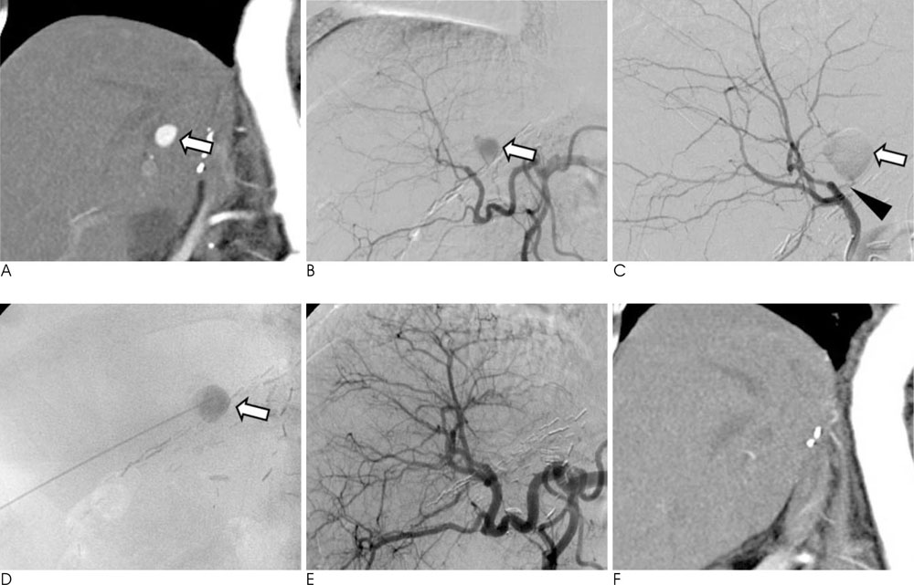
Fig. 2 A. Arterial phase, coronal reformatting CT shows a well-enhancing intrahepatic pseudoaneurysm (arrow). B, C. Hepatic arteriograms show that the pseudoaneurysm (arrow) arises from a small branch (arrowhead) of the posterosuperior intrahepatic artery. D. Percutaneous puncture of the pseudoaneurysm (arrow) was performed. E. The final angiogram shows disappearance of the pseudoaneurysm. Note the relatively well-preserved intrahepatic arterial branches. F. Coronal reformatting CT obtained 1 week after embolization shows that the pseudoaneurysm has disappeared.

Fig. 3 A. Contrast-enhanced CT shows a pseudoaneurysm (arrow) in the graft liver. B, C. Four- (B) and 18-day (C) follow-up CT images show gradual thrombosis of the pseudoaneurysm (arrow
Reference
-
1. Kim KR, Ko GY, Sung KB, Yoon HK, Shin JH, Song HY, et al. Transjugular liver biopsy in patients with living donor liver transplantation: comparison with percutaneous biopsy. Liver Transpl. 2008; 14:971–979.2. Kalambokis G, Manousou P, Vibhakorn S, Marelli L, Cholongitas E, Senzolo M, et al. Transjugular liver biopsy-Indications, adequacy, quality of specimens, and complications - a systematic review. J Hepatol. 2007; 47:284–294.3. Kaye R, Sane SS, Towbin RB. Pediatric intervention: an update-part II. J Vasc Interv Radiol. 2000; 11:807–822.4. Roche CJ, Lee WK, Duddalwar VA, Nicolaou S, Munk PL, Morris DC. Intrahepatic pseudoaneurysm complicating transjugular biopsy of the liver. AJR Am J Roentgenol. 2001; 177:819–821.5. Dillon BJ, Alomari AI. Delayed formation and rupture of a pseudoaneurysm after Transjugular liver biopsy in a pediatric bone marrow transplant patient: imaging and endovascular treatment. Cardiovasc Intervent Radiol. 2009; 32:377–380.6. Schuster R, Broumandi DD, Lee AA, Waxman K. Percutaneous thrombin injection to treat a post-traumatic hepatic pseudoaneurysm. J Trauma. 2005; 59:1022–1024.7. Esposito AA, Nicolini A, Meregaglia D, Sangiovanni A, Biondetti P. Role of transjugular liver biopsy in the diagnosis and therapeutic management of patients with severe liver disease. Radiol Med. 2008; 113:1008–1017.8. Gurakuqi GC, Stadlbauer V, Portugaller HR, Hogenauer C, Trauner M, Stauber RE. Fatal hemobilia resulting from an iatrogenic arteriobiliary fistula as a rare complication of transjugular liver biopsy. Eur J Gastroenterol Hepatol. 2008; 20:83–86.9. Merhav H, Zajko AB, Dodd GD, Pinna A. Percutaneous transhepatic embolization of an intrahepatic pseudoaneurysm following liver biopsy in a liver transplant patient. Transpl Int. 1993; 6:239–241.10. Hanson JM, Atri M, Power N. Ultrasound-guided thrombin injection of iatrogenic groin pseudoaneurysm: doppler features and technical tips. Br J Radiol. 2008; 81:154–163.
- Full Text Links
-
- Actions
-
Cited
- CITED
-
- Close
- Share
-
- Similar articles
-
- Treating systemic inflammation by transjugular intrahepatic portosystemic shunt: Editorial on “Insertion of a transjugular intrahepatic portosystemic shunt leads to sustained reversal of systemic inflammation in patients with decompensated liver cirrhosis”
- Decreasing systemic inflammation after TIPS: Still hope for the liver: Reply to correspondence on “Insertion of a transjugular intrahepatic portosystemic shunt leads to sustained reversal of systemic inflammation in patients with decompensated liver cirrhosis”
- Two Cases of Percutaneous Transhepatic Choledochoscopy Treatment of Intrahepatic Duct Stones that Occurred after Living Donor Liver Transplantation
- A case of probable endotipsitis after transjugular intrahepatic portasystemic shunt
- Rescue therapy for bleeding ectopic ileal varices with a transjugular intrahepatic portosystemic shunt and antegrade variceal embolization
