Acetabular Subchondral Bone Decortication and Its Role in the Outcome of Cemented Total Hip Replacement in Young Patients
- Affiliations
-
- 1Department of Trauma and Orthopaedic Surgery, Heart of England NHS Foundation Trust, Birmingham, United Kingdom. kanaigarala@gmail.com
- 2Department of Trauma of Orthopaedic Surgery, University Hospitals Coventry and Warwickshire, Coventry, United Kingdom.
- 3Department of Radiology, St Thomas Hospital, London, United Kingdom.
- KMID: 2419514
- DOI: http://doi.org/10.5371/hp.2018.30.3.182
Abstract
- PURPOSE
Long-term fixation of cemented acetabular components can be problematic in younger active patients. Our technique is put forward to improve outcomes and maximize implant survivorship in this particular patient population.
MATERIALS AND METHODS
We report on a cohort of young adult patients (less than 55 years old) with cemented total hip replacement (THR) using a novel technique in preparing and cementing the acetabulum with a minimum follow-up of 10 years (mean follow-up, 14 years). Retrospectively collected data on clinical and radiological outcomes were reviewed.
RESULTS
Sixty-five THRs were performed with the minimum study follow-up period. Average age for patients was 44 years old (range, 19-55 years). The mean Hip Disability and Osteoarthritis Outcome Score for patients at final appointment was 92.7. Radiographs taken at an average of 14 years after operation showed 63 of 65 hips showed no evidence of any radiological loosening. Cup survivorship was 100% at the end of the study period.
CONCLUSION
Our technique of preparing the acetabulum in combination with cement fixation is reproducible with excellent results in a cohort of patients prone to early aseptic loosening of the acetabular component.
Keyword
MeSH Terms
Figure
-

Fig. 1 The true floor of the acetabulum is 1st exposed with a gouge osteotome.

Fig. 2 The floor is fully exposed with a reamer.
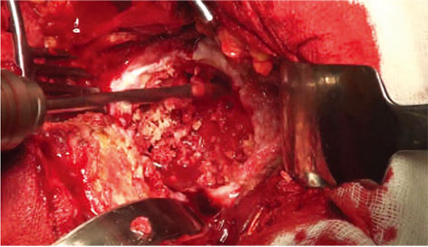
Fig. 3 The subchondral bone is prepared with a 6-mm drill to begin to expose the bone under the subchondral sclerotic plate.

Fig. 4 The subchondral bone plate is further exposed by skimming the top layer of sclerotic bone off with the gouge osteotome.
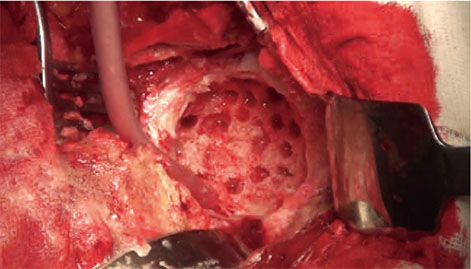
Fig. 5 A figure of the finished acetabular preparation, just prior to cementation.
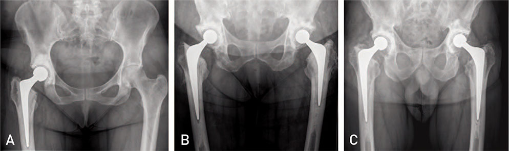
Fig. 6 Post-operative radiographs of four patients having gone total hip replacement with subchondral bone decortication. (A) Sixteen years and (B, C) seventeen years after operaion.

Fig. 7 Pie charts showing responses to satisfaction questionnaires post hip replacement at the latest follow-up for each question (mean 14 years).
Reference
-
1. Clarke A, Pulikottil-Jacob R, Grove A, et al. Total hip replacement and surface replacement for the treatment of pain and disability resulting from end-stage arthritis of the hip (review of technology appraisal guidance 2 and 44): systematic review and economic evaluation. Health Technol Assess. 2015; 19:1–668. vii–viii.
Article2. Malchau H, Herberts P, Ahnfelt L. Prognosis of total hip replacement in Sweden. Follow-up of 9,675 operations performed 1978-1990. Acta Orthop Scand. 1993; 64:497–506.
Article3. Timperley AJ, Whitehouse SL, Hourigan PG. The influence of a suction device on fixation of a cemented cup using RSA. Clin Orthop Relat Res. 2009; 467:792–798.
Article4. Havelin LI, Fenstad AM, Salomonsson R, et al. The Nordic Arthroplasty Register Association: a unique collaboration between 3 national hip arthroplasty registries with 280, 201 THRs. Acta Orthop. 2009; 80:393–401.5. Kynaston-Pearson F, Ashmore AM, Malak TT, et al. Primary hip replacement prostheses and their evidence base: systematic review of literature. BMJ. 2013; 347:f6956.
Article6. Burston BJ, Yates PJ, Hook S, Moulder E, Whitley E, Bannister GC. Cemented polished tapered stems in patients less than 50 years of age: a minimum 10-year follow-up. J Arthroplasty. 2010; 25:692–699.
Article7. Berry DJ, Harmsen WS, Cabanela ME, Morrey BF. Twenty-five-year survivorship of two thousand consecutive primary Charnley total hip replacements: factors affecting survivorship of acetabular and femoral components. J Bone Joint Surg Am. 2002; 84-A:171–177.8. Callaghan JJ, Albright JC, Goetz DD, Olejniczak JP, Johnston RC. Charnley total hip arthroplasty with cement. Minimum twenty-five-year follow-up. J Bone Joint Surg Am. 2000; 82:487–497.9. Ballard WT, Callaghan JJ, Sullivan PM, Johnston RC. The results of improved cementing techniques for total hip arthroplasty in patients less than fifty years old. A ten-year follow-up study. J Bone Joint Surg Am. 1994; 76:959–964.
Article10. Aspenberg P, Van der Vis H. Migration, particles, and fluid pressure. A discussion of causes of prosthetic loosening. Clin Orthop Relat Res. 1998; (352):75–80.11. Mjöberg B. The theory of early loosening of hip prostheses. Orthopedics. 1997; 20:1169–1175.
Article12. Flivik G, Sanfridsson J, Onnerfält R, Kesteris U, Ryd L. Migration of the acetabular component: effect of cement pressurization and significance of early radiolucency: a randomized 5-year study using radiostereometry. Acta Orthop. 2005; 76:159–168.
Article13. Majkowski RS, Bannister GC, Miles AW. The effect of bleeding on the cement-bone interface. An experimental study. Clin Orthop Relat Res. 1994; (299):293–297.14. Carter DR, Vasu R, Harris WH. Periacetabular stress distributions after joint replacement with subchondral bone retention. Acta Orthop Scand. 1983; 54:29–35.
Article15. Vasu R, Carter DR, Harris WH. Stress distributions in the acetabular region--I. Before and after total joint replacement. J Biomech. 1982; 15:155–164.
Article16. Ritter MA, Thong AE. The role of cemented sockets in 2004: is there one? J Arthroplasty. 2004; 19:4 Suppl 1. 92–94.17. Sutherland AG, D'Arcy S, Smart D, Ashcroft GP. Removal of the subchondral plate in acetabular preparation. Int Orthop. 2000; 24:19–22.
Article18. Crites BM, Berend ME, Ritter MA. Technical considerations of cemented acetabular components: a 30-year evaluation. Clin Orthop Relat Res. 2000; (381):114–119.19. Mulroy WF, Harris WH. Acetabular and femoral fixation 15 years after cemented total hip surgery. Clin Orthop Relat Res. 1997; (337):118–128.
Article20. Pedersen AB, Mehnert F, Havelin LI, et al. Association between fixation technique and revision risk in total hip arthroplasty patients younger than 55 years of age. Results from the Nordic Arthroplasty Register Association. Osteoarthritis Cartilage. 2014; 22:659–667.
Article21. Eskelinen A, Remes V, Helenius I, Pulkkinen P, Nevalainen J, Paavolainen P. Uncemented total hip arthroplasty for primary osteoarthritis in young patients: a mid-to long-term follow-up study from the Finnish Arthroplasty Register. Acta Orthop. 2006; 77:57–70.
Article22. Mäkelä KT, Eskelinen A, Pulkkinen P, Paavolainen P, Remes V. Results of 3,668 primary total hip replacements for primary osteoarthritis in patients under the age of 55 years: A follow-up of a previous report from the Finnish Arthroplasty Register. Acta Orthop. 2011; 82:521–529.
Article23. Jameson SS, Baker PN, Mason J, et al. The design of the acetabular component and size of the femoral head influence the risk of revision following 34 721 single-brand cemented hip replacements: a retrospective cohort study of medium-term data from a National Joint Registry. J Bone Joint Surg Br. 2012; 94:1611–1617.24. Culliford DJ, Maskell J, Beard DJ, Murray DW, Price AJ, Arden NK. Temporal trends in hip and knee replacement in the United Kingdom: 1991 to 2006. J Bone Joint Surg Br. 2010; 92:130–135.25. Streit MR, Weiss S, Andreas F, et al. 10-year results of the uncemented Allofit press-fit cup in young patients: 121 hips followed for 10-12 years. Acta Orthop. 2014; 85:368–374.
Article26. Flivik G, Kristiansson I, Ryd L. Positive effect of removal of subchondral bone plate for cemented acetabular component fixation in total hip arthroplasty: a randomised RSA study with ten-year follow-up. Bone Joint J. 2015; 97-B:35–44.27. Nilsdotter A, Bremander A. Measures of hip function and symptoms: Harris Hip Score (HHS), Hip Disability and Osteoarthritis Outcome Score (HOOS), Oxford Hip Score (OHS), Lequesne Index of Severity for Osteoarthritis of the Hip (LISOH), and American Academy of Orthopedic Surgeons (AAOS) Hip and Knee Questionnaire. Arthritis Care Res (Hoboken). 2011; 63:Suppl 11. S200–S207.28. Nilsdotter AK, Lohmander LS, Klässbo M, Roos EM. Hip disability and osteoarthritis outcome score (HOOS)--validity and responsiveness in total hip replacement. BMC Musculoskelet Disord. 2003; 4:10.
Article29. Clement ND, Biant LC, Breusch SJ. Total hip arthroplasty: to cement or not to cement the acetabular socket? A critical review of the literature. Arch Orthop Trauma Surg. 2012; 132:411–427.
Article30. National Joint Registry (NJR). 14th Annual Report of the NJR for England, Wales, Northern Ireland and the Isle of Man [Internet]. Hemel Hempstead, UK: NJR;2017. 2018 jul 1. Available from: http://www.njrcentre.org.uk/njrcentre/NewsandEvents/NJR14thAnnualReportrecordnumberofproceduresduring201617/tabid/1453/Default.aspx.31. Wroblewski BM, Siney PD, Fleming PA. Wear of the cup in the Charnley LFA in the young patient. J Bone Joint Surg Br. 2004; 86:498–503.
Article32. Mulroy RD Jr, Harris WH. The effect of improved cementing techniques on component loosening in total hip replacement. An 11-year radiographic review. J Bone Joint Surg Br. 1990; 72:757–760.
Article33. Schmalzried TP, Kwong LM, Jasty M, et al. The mechanism of loosening of cemented acetabular components in total hip arthroplasty. Analysis of specimens retrieved at autopsy. Clin Orthop Relat Res. 1992; (274):60–78.34. Hultmark P, Höstner J, Herberts P, Kärrholm J. Radiographic evaluation of Charnley cups used in first-time revision: repeated observations for 7-15 years. J Arthroplasty. 2003; 18:1005–1015.35. Ritter MA, Zhou H, Keating CM, et al. Radiological factors influencing femoral and acetabular failure in cemented Charnley total hip arthroplasties. J Bone Joint Surg Br. 1999; 81:982–986.
Article36. García-Cimbrelo E, Diez-Vazquez V, Madero R, Munuera L. Progression of radiolucent lines adjacent to the acetabular component and factors influencing migration after Charnley low-friction total hip arthroplasty. J Bone Joint Surg Am. 1997; 79:1373–1380.
