Ann Dermatol.
2018 Jun;30(3):373-375. 10.5021/ad.2018.30.3.373.
A Case of Plexiform Fibrohistiocytic Tumor on Finger
- Affiliations
-
- 1Department of Dermatology, Asan Medical Center, University of Ulsan College of Medicine, Seoul, Korea. uucm79@gmail.com
- KMID: 2419187
- DOI: http://doi.org/10.5021/ad.2018.30.3.373
Abstract
- No abstract available.
MeSH Terms
Figure
-
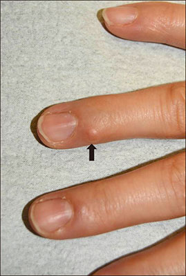
Fig. 1 A solitary, 4×4 mm, firm, nontender, skin-colored, round-shaped nodule on the right fourth finger (indicated by a black arrow).
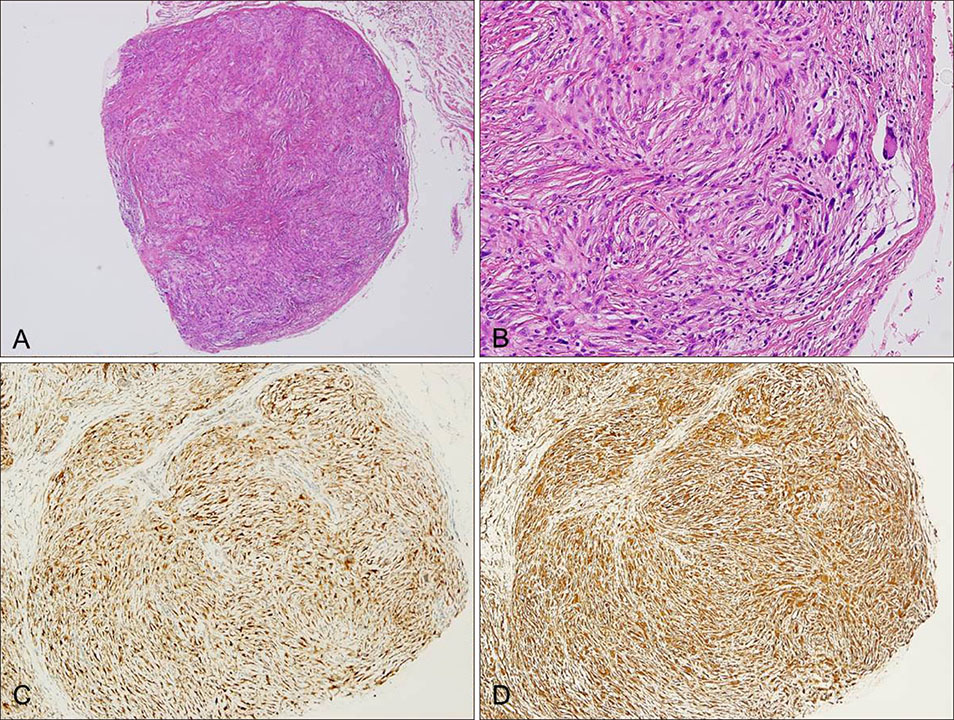
Fig. 2 (A) A relatively well-demarcated dermal tumor mass showing nodular and plexiform growth pattern (H&E, ×40). (B) Peripheral osteoclast-like giant cells and foamy clear cells of histiocytic type surrounded by fibroblast-like spindle cells showing a characteristic biphasic pattern without significant cytologic atypia and pleomorphism (H&E, ×200). (C, D) Immunopositivity for (C) CD68 and (D) vimentin (×200).
Reference
-
1. Cho S, Chang SE, Choi JH, Sung KJ, Moon KC, Koh JK. Myxoid plexiform fibrohistiocytic tumour. J Eur Acad Dermatol Venereol. 2002; 16:519–521.
Article2. Enzinger FM, Zhang RY. Plexiform fibrohistiocytic tumor presenting in children and young adults. An analysis of 65 cases. Am J Surg Pathol. 1988; 12:818–826.
Article3. Moosavi C, Jha P, Fanburg-Smith JC. An update on plexiform fibrohistiocytic tumor and addition of 66 new cases from the Armed Forces Institute of Pathology, in honor of Franz M. Enzinger, MD. Ann Diagn Pathol. 2007; 11:313–319.
Article4. Taher A, Pushpanathan C. Plexiform fibrohistiocytic tumor: a brief review. Arch Pathol Lab Med. 2007; 131:1135–1138.
Article5. Shido K, Fujimura T, Kakizaki A, Furudate S, Asano M, Aiba S. Plexiform fibrohistiocytic tumor on the ear: case report and immunohistochemical investigation of stromal factor. Case Rep Dermatol. 2016; 8:26–30.
Article
