Computed Diffusion-Weighted Imaging in Prostate Cancer: Basics, Advantages, Cautions, and Future Prospects
- Affiliations
-
- 1Department of Radiology, Kobe University Graduate School of Medicine, Kobe 650-0017, Japan. yoshiu@med.kobe-u.ac.jp
- 2Department of Radiology, Kawasaki Medical School, Kurashiki 701-0192, Japan.
- 3Advanced Biomedical Imaging Research Center, Kobe University Graduate School of Medicine, Kobe 650-0017, Japan.
- 4Department of Urology, Kobe University Graduate School of Medicine, Kobe 650-0017, Japan.
- KMID: 2418545
- DOI: http://doi.org/10.3348/kjr.2018.19.5.832
Abstract
- Computed diffusion-weighted MRI is a recently proposed post-processing technique that produces b-value images from diffusion-weighted imaging (DWI), acquired using at least two different b-values. This article presents an argument for computed DWI for prostate cancer by viewing four aspects of DWI: fundamentals, image quality and diagnostic performance, computing procedures, and future uses.
Keyword
Figure
-
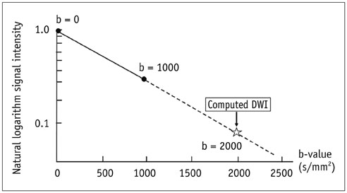
Fig. 1 Schema of computed DWI. For example, DW images at b-values of 0 s/mm2 and 1000 s/mm2 can be used for computation of DW images at b-value of 2000 s/mm2 using estimated apparent diffusion coefficient values based on mono-exponential model. DWI = diffusion weighted imaging
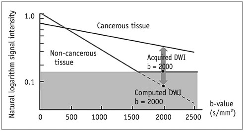
Fig. 2 Theory of advantage of computed DWI. Computed DWI potentially suppresses background noise, maintaining lesion signal and leading to better contrast of cancerous lesion.
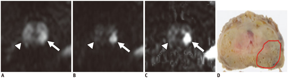
Fig. 3 Computed DWI of prostate cancer. With acquired DWI at b = 1000 s/mm2 (A), bilateral lobes of PZ show abnormal signal intensity (arrow and arrowhead). With both acquired DWI at b = 1000 s/mm2 (B) and computed DWI at b = 2000 s/mm2 (C), left lobe of PZ shows abnormal signal intensity (arrows), whereas right lobe of PZ shows normal signal (arrowheads). Pathological specimen confirmed prostate cancer with GS 4 + 3 in left side of PZ (delineated by solid line, D) and no cancer in right side of PZ. PZ = peripheral zone
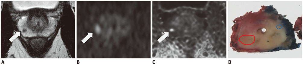
Fig. 4 High spatial-resolution computed DWI. Abnormal signal in right side of PZ is shown on T2WI (arrow, A). With normal resolution DWI at b = 2000 s/mm2 (arrow, B), abnormal signal in right side of PZ is depicted but is distorted compared to T2WI. With high resolution computed DWI at b = 2000 s/mm2 (arrow, C), abnormal signal in right side of PZ is shown clearly and less distorted. In this image, boundary of prostate is also depicted. Pathological specimen revealed cancer with GS 3 + 4 in right side of PZ (delineated by red line, D). Although there was small tumor with GS 3 + 3 on left side of PZ, it was not depicted on magnetic resonance imaging (delineated by blue line, D). T2WI = T2-weighted imaging
Reference
-
1. Koh DM, Collins DJ. Diffusion-weighted MRI in the body: applications and challenges in oncology. AJR Am J Roentgenol. 2007; 188:1622–1635.
Article2. Kim CK, Park SY, Park JJ, Park BK. Diffusion-weighted MRI as a predictor of extracapsular extension in prostate cancer. AJR Am J Roentgenol. 2014; 202:W270–W276.
Article3. Chong Y, Kim CK, Park SY, Park BK, Kwon GY, Park JJ. Value of diffusion-weighted imaging at 3 T for prediction of extracapsular extension in patients with prostate cancer: a preliminary study. AJR Am J Roentgenol. 2014; 202:772–777.
Article4. Hoeks CM, Barentsz JO, Hambrock T, Yakar D, Somford DM, Heijmink SW, et al. Prostate cancer: multiparametric MR imaging for detection, localization, and staging. Radiology. 2011; 261:46–66.
Article5. Tamada T, Sone T, Jo Y, Yamamoto A, Ito K. Diffusion-weighted MRI and its role in prostate cancer. NMR Biomed. 2014; 27:25–38.
Article6. Metens T, Miranda D, Absil J, Matos C. What is the optimal b value in diffusion-weighted MR imaging to depict prostate cancer at 3T? Eur Radiol. 2012; 22:703–709.
Article7. Katahira K, Takahara T, Kwee TC, Oda S, Suzuki Y, Morishita S, et al. Ultra-high-b-value diffusion-weighted MR imaging for the detection of prostate cancer: evaluation in 201 cases with histopathological correlation. Eur Radiol. 2011; 21:188–196.
Article8. Ueno Y, Kitajima K, Sugimura K, Kawakami F, Miyake H, Obara M, et al. Ultra-high b-value diffusion-weighted MRI for the detection of prostate cancer with 3-T MRI. J Magn Reson Imaging. 2013; 38:154–160.
Article9. Tamada T, Kanomata N, Sone T, Jo Y, Miyaji Y, Higashi H, et al. High b value (2,000 s/mm2) diffusion-weighted magnetic resonance imaging in prostate cancer at 3 Tesla: comparison with 1,000 s/mm2 for tumor conspicuity and discrimination of aggressiveness. PLoS One. 2014; 9:e96619.10. Koh DM, Blackledge M, Padhani AR, Takahara T, Kwee TC, Leach MO, et al. Whole-body diffusion-weighted MRI: tips, tricks, and pitfalls. AJR Am J Roentgenol. 2012; 199:252–262.
Article11. Blackledge MD, Leach MO, Collins DJ, Koh DM. Computed diffusion-weighted MR imaging may improve tumor detection. Radiology. 2011; 261:573–581.
Article12. Rohde GK, Barnett AS, Basser PJ, Marenco S, Pierpaoli C. Comprehensive approach for correction of motion and distortion in diffusion-weighted MRI. Magn Reson Med. 2004; 51:103–114.
Article13. Rosenkrantz AB, Chandarana H, Hindman N, Deng FM, Babb JS, Taneja SS, et al. Computed diffusion-weighted imaging of the prostate at 3 T: impact on image quality and tumour detection. Eur Radiol. 2013; 23:3170–3177.
Article14. Bammer R, Keeling SL, Augustin M, Pruessmann KP, Wolf R, Stollberger R, et al. Improved diffusion-weighted single-shot echo-planar imaging (EPI) in stroke using sensitivity encoding (SENSE). Magn Reson Med. 2001; 46:548–554.
Article15. Zhu Y. Parallel excitation with an array of transmit coils. Magn Reson Med. 2004; 51:775–784.
Article16. Maas MC, Fütterer JJ, Scheenen TW. Quantitative evaluation of computed high B value diffusion-weighted magnetic resonance imaging of the prostate. Invest Radiol. 2013; 48:779–786.
Article17. Ueno Y, Takahashi S, Kitajima K, Kimura T, Aoki I, Kawakami F, et al. Computed diffusion-weighted imaging using 3-T magnetic resonance imaging for prostate cancer diagnosis. Eur Radiol. 2013; 23:3509–3516.
Article18. Yoshida R, Yoshizako T, Katsube T, Tamaki Y, Ishikawa N, Kitagaki H. Computed diffusion-weighted imaging using 1.5-T magnetic resonance imaging for prostate cancer diagnosis. Clin Imaging. 2017; 41:78–82.
Article19. Lim HK, Kim JK, Kim KA, Cho KS. Prostate cancer: apparent diffusion coefficient map with T2-weighted images for detection--a multireader study. Radiology. 2009; 250:145–151.
Article20. Haider MA, van der Kwast TH, Tanguay J, Evans AJ, Hashmi AT, Lockwood G, et al. Combined T2-weighted and diffusion-weighted MRI for localization of prostate cancer. AJR Am J Roentgenol. 2007; 189:323–328.
Article21. Kim CK, Park BK, Kim B. High-b-value diffusion-weighted imaging at 3 T to detect prostate cancer: comparisons between b values of 1,000 and 2,000 s/mm2. AJR Am J Roentgenol. 2010; 194:W33–W37.22. Kwon MR, Kim CK, Kim JH. PI-RADS version 2: evaluation of diffusion-weighted imaging interpretation between b = 1000 and b = 1500 s mm−2. Br J Radiol. 2017; 90:2017043.23. Koo JH, Kim CK, Choi D, Park BK, Kwon GY, Kim B. Diffusion-weighted magnetic resonance imaging for the evaluation of prostate cancer: optimal B value at 3T. Korean J Radiol. 2013; 14:61–69.
Article24. Weinreb JC, Barentsz JO, Choyke PL, Cornud F, Haider MA, Macura KJ, et al. PI-RADS prostate imaging - Reporting and data system: 2015, Version 2. Eur Urol. 2016; 69:16–40.
Article25. Vargas HA, Hötker AM, Goldman DA, Moskowitz CS, Gondo T, Matsumoto K, et al. Updated prostate imaging reporting and data system (PIRADS v2) recommendations for the detection of clinically significant prostate cancer using multiparametric MRI: critical evaluation using whole-mount pathology as standard of reference. Eur Radiol. 2016; 26:1606–1612.
Article26. Ueno Y, Takahashi S, Ohno Y, Kitajima K, Yui M, Kassai Y, et al. Computed diffusion-weighted MRI for prostate cancer detection: the influence of the combinations of b-values. Br J Radiol. 2015; 88:20140738.27. Koh DM, Collins DJ, Orton MR. Intravoxel incoherent motion in body diffusion-weighted MRI: reality and challenges. AJR Am J Roentgenol. 2011; 196:1351–1361.
Article28. Ogura A, Koyama D, Hayashi N, Hatano I, Osakabe K, Yamaguchi N. Optimal b values for generation of computed high-b-value DW images. AJR Am J Roentgenol. 2016; 206:713–718.
Article29. Rosenkrantz AB, Parikh N, Kierans AS, Kong MX, Babb JS, Taneja SS, et al. Prostate cancer detection using computed very high b-value diffusion-weighted imaging: how high should we go? Acad Radiol. 2016; 23:704–711.
Article30. Kimura T, Machii Y. Computed diffusion weighted imaging under Rician noise distribution. Abstract 3574. In : Proceedings of the International Society for Magnetic Resonance in Medicine; May 4–11, 2012; Melbourne, AU. International Society for Magnetic Resonance in Medicine.31. Verma S, Sarkar S, Young J, Venkataraman R, Yang X, Bhavsar A, et al. Evaluation of the impact of computed high b-value diffusion-weighted imaging on prostate cancer detection. Abdom Radiol (NY). 2016; 41:934–945.
Article32. Medved M, Soylu-Boy FN, Karademir I, Sethi I, Yousuf A, Karczmar GS, et al. High-resolution diffusion-weighted imaging of the prostate. AJR Am J Roentgenol. 2014; 203:85–90.
Article33. Hwang J, Hong SS, Kim HJ, Chang YW, Nam BD, Oh E, et al. Reduced field-of-view diffusion-weighted MRI in patients with cervical cancer. Br J Radiol. 2018; 91:20170864.
Article34. Bittencourt LK, Attenberger UI, Lima D, Strecker R, de Oliveira A, Schoenberg SO, et al. Feasibility study of computed vs measured high b-value (1400 s/mm2) diffusion-weighted MR images of the prostate. World J Radiol. 2014; 6:374–380.35. Ueno Y, Takahashi S, Ohno Y, Kyotani K, Yui M, kassai Y, et al. High-resolution computed DWI with high b-value: a preliminary study for improving prostate cancer detection at 3T MR system. Abstract 1540. In : Proceedings of the International Society for Magnetic Resonance in Medicine; May 30–June 5, 2015; Toronto, ON, Canada. International Society for Magnetic Resonance in Medicine.36. Hambrock T, Somford DM, Huisman HJ, van Oort IM, Witjes JA, Hulsbergen-van de Kaa CA, et al. Relationship between apparent diffusion coefficients at 3.0-T MR imaging and Gleason grade in peripheral zone prostate cancer. Radiology. 2011; 259:453–461.
Article37. Waseda Y, Yoshida S, Takahara T, Kwee TC, Matsuoka Y, Saito K, et al. Utility of computed diffusion-weighted MRI for predicting aggressiveness of prostate cancer. J Magn Reson Imaging. 2017; 46:490–496.
Article
- Full Text Links
-
- Actions
-
Cited
- CITED
-
- Close
- Share
-
- Similar articles
-
- RE: Diffusion-Weighted Imaging of Prostate Cancer: How Can We Use It Accurately?
- Multidisciplinary Functional MR Imaging for Prostate Cancer
- Basics for Pediatric Brain Tumor Imaging: Techniques and Protocol Recommendations
- Effectiveness of Bi-Parametric MR/US Fusion Biopsy for Detecting Clinically Significant Prostate Cancer in Prostate Biopsy Naïve Men
- Comparison of Computed Diffusion-Weighted Imaging b2000 and Acquired Diffusion-Weighted Imaging b2000 for Detection of Prostate Cancer
