Symptomatic Anomalous Coronary Artery Origin Diagnosis and Interventions
- Affiliations
-
- 1Department of Cardiology, Rovigo General Hospital, Rovigo, Italy. jackyheart@libero.it
- KMID: 2414916
- DOI: http://doi.org/10.4070/kcj.2018.0025
Abstract
- No abstract available.
MeSH Terms
Figure
-

Figure 1 Subselective right coronary angiography demonstrating an anomalous origin (asterisk) of the RCA from the left coronary sinus of valsalva on anteroposterior view. RCA = right coronary artery.

Figure 2 A CT angiographic scan demonstrate an anomalous origin (asterisk) of RCA (A) without a clear demonstration of the intramural course of the vessel (B). CT = computed tomography; RCA = right coronary artery.

Figure 3 Transthoracic echocardiogram in parasternal short axis view shows an intramural decourse (asterisk) of the of the proximal portion of RCA (A, B: the red conturns highlight the borders of the aortic annulus and the intramural wall of the vessel). AW = atrioventricular valve; AV = aortic valve; CAW = common atrioventricular valve; RCA = right coronary artery.
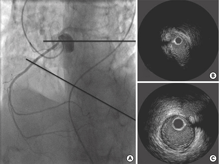
Figure 4 Urgent coronary angiography in right anterior oblique projection (A) and IVUS examination: a compression of the intramural course is apparent being the proximal part of the vessel squeezed into an elliptical shape with clearly reduced luminal area and no real plaque burden (B). The rest of the vessel is free from significant atherosclerosis (C). IVUS = intravascular ultrasound.

Figure 5 IVUS control after successful stenting (A): the first 20 mm of the proximal portion of the vessel, that is the length of the intramural course were covered by the stent with a normal luminal area (B, C). IVUS = intravascular ultrasound.
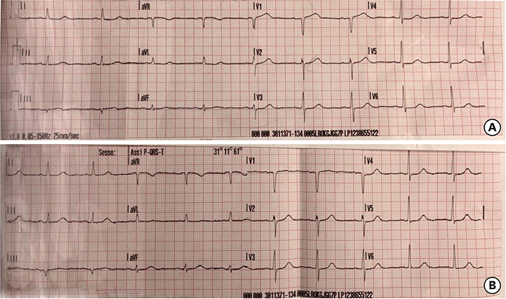
Figure 6 Electrocardiogram (A) pre- and (B) post-successful stenting of the intramural course demonstrating resolution of the ST changes in inferior leads, in particular D3.
Reference
-
1. Angelini P, Uribe C, Monge J, Tobis JM, Elayda MA, Willerson JT. Origin of the right coronary artery from the opposite sinus of Valsalva in adults: characterization by intravascular ultrasonography at baseline and after stent angioplasty. Catheter Cardiovasc Interv. 2015; 86:199–208.
Article2. Rigatelli G, Cardaioli P. Endovascular therapy for congenital coronary artery anomalies in adults. J Cardiovasc Med (Hagerstown). 2008; 9:113–121.
Article
- Full Text Links
-
- Actions
-
Cited
- CITED
-
- Close
- Share
-
- Similar articles
-
- Two Cases of Anomalous Origin of Coronary Artery
- Anomalous Origin of Left Main Coronary Artery from the Right Coronary Artery: Echocardiographic Diagnosis
- Anomalous origin of the left coronary artery from the pulmonary artery
- Sudden Death Associated with Anomalous Left Coronary Artery Origin from Right Sinus of Valsalva with Posterior Course
- Role of Transesophageal Echocardiography in Identifying Anomalous Origin and Course of Coronary Arteries
