Clin Endosc.
2018 May;51(3):274-278. 10.5946/ce.2017.098.
Capability of Radial- and Convex-Arrayed Echoendoscopes for Visualization of the Pancreatobiliary Junction
- Kanno Y

- Ito K
- Koshita S
- Ogawa T
- Kusunose H
- Masu K
- Sakai T
- Murabayashi T
- Hasegawa S
- Kozakai F
- Kawakami Y
- Fujii Y
- Noda Y
- Affiliations
-
- 1Sendai City Medical Center, Sendai, Japan.
- KMID: 2414869
- DOI: http://doi.org/10.5946/ce.2017.098
Abstract
- BACKGROUND/AIMS
Although both radial- and convex-arrayed endoscopic ultrasonography (EUS) scopes are widely used for observational EUS examinations, there have been few comparative studies on their power of visualization. The aim of this study was to evaluate the capability of these EUS scopes for observation of the pancreatobiliary junction.
METHODS
The rate of successful visualization of the pancreatobiliary junction was retrospectively compared between a radial-arrayed and a convex-arrayed echoendoscope, from a prospectively maintained database. Study periods were defined as January 2010 to December 2012 for the radial group, and February 2015 to October 2016 for the convex group because the respective scope was mainly used during those periods.
RESULTS
During the study period, 1,660 cases with radial EUS and 1,984 cases with convex EUS were recruited. The success rates of observation of the pancreatobiliary junction were 80.0% and 89.5%, respectively (p < 0.0001).
CONCLUSIONS
The capability of visualization of the pancreatobiliary junction in observational EUS was found to be better with a convex-arrayed than with a radial-arrayed echoendoscope.
Figure
-
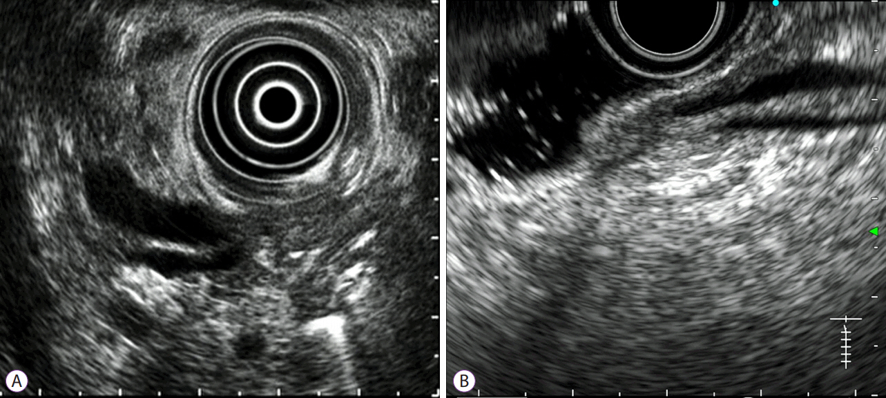
Fig. 1. “Well observed” endoscopic ultrasonography (EUS) views of the pancreatobiliary junction. “Well observed” was defined as a case in which the distal ends of both the bile duct and the main pancreatic duct were clearly observed with awareness of positional relation between these ducts, the duodenal ampulla, and the duodenal muscular layer. It does not require visualization of both the ducts in one image at the same time, especially with convex EUS. (A) View by a radial-arrayed echoendoscope. (B) View by a convex-arrayed echoendoscope.
Reference
-
1. Sugiyama M, Atomi Y. Endoscopic ultrasonography for diagnosing choledocholithiasis: a prospective comparative study with ultrasonography and computed tomography. Gastrointest Endosc. 1997; 45:143–146.
Article2. Kondo S, Isayama H, Akahane M, et al. Detection of common bile duct stones: comparison between endoscopic ultrasonography, magnetic resonance cholangiography, and helical-computed-tomographic cholangiography. Eur J Radiol. 2005; 54:271–275.
Article3. Mirbagheri SA, Mohamadnejad M, Nasiri J, Vahid AA, Ghadimi R, Malekzadeh R. Prospective evaluation of endoscopic ultrasonography in the diagnosis of biliary microlithiasis in patients with normal transabdominal ultrasonography. J Gastrointest Surg. 2005; 9:961–964.
Article4. Ney MV, Maluf-Filho F, Sakai P, Zilberstein B, Gama-Rodrigues J, Rosa H. Echo-endoscopy versus endoscopic retrograde cholangiography for the diagnosis of choledocholithiasis: the influence of the size of the stone and diameter of the common bile duct. Arq Gastroenterol. 2005; 42:239–243.5. Mitake M, Nakazawa S, Naitoh Y, et al. Value of endoscopic ultrasonography in the detection of anomalous connections of the pancreatobiliary duct. Endoscopy. 1991; 23:117–120.
Article6. Sugiyama M, Atomi Y. Endoscopic ultrasonography for diagnosing anomalous pancreaticobiliary junction. Gastrointest Endosc. 1997; 45:261–267.
Article7. Tanaka M, Fernández-del Castillo C, Adsay V, et al. International consensus guidelines 2012 for the management of IPMN and MCN of the pancreas. Pancreatology. 2012; 12:183–197.
Article8. Katanuma A, Maguchi H, Osanai M, Takahashi K. The difference in the capability of delineation between convex and radial arrayed echoendoscope for pancreas and biliary tract; case reports from the standpoint of both convex and radial arrayed echoendoscope. Dig Endosc. 2011; 23 Suppl 1:2–8.
Article9. Hara K, Bhatia V, Hijioka S, Mizuno N, Yamao K. A convex EUS is useful to diagnose vascular invasion of cancer, especially hepatic hilus cancer. Dig Endosc. 2011; 23 Suppl 1:26–28.
Article10. Kaneko M, Katanuma A, Maguchi H, et al. Prospective, randomized, comparative study of delineation capability of radial scanning and curved linear array endoscopic ultrasound for the pancreaticobiliary region. Endosc Int Open. 2014; 2:E160–E170.
Article11. Ito K, Fujita N, Noda Y, et al. Preoperative evaluation of ampullary neoplasm with EUS and transpapillary intraductal US: a prospective and histopathologically controlled study. Gastrointest Endosc. 2007; 66:740–747.
Article
- Full Text Links
-
- Actions
-
Cited
- CITED
-
- Close
- Share
-
- Similar articles
-
- Convex versus Radial Echoendoscopes - Comparison of Capability for Evaluating the Pancreatobiliary Junction
- Evaluation of the Feasibility and Efficacy of Forward-Viewing Endoscopic Ultrasound
- Clinical Observation for the Antegrade Pyelograghy
- Process and Renewal of Pancreatobiliary Cerification System
- Recent Updates of Immunoglobulin G4-related Pancreatobiliary Disease
