Ann Dermatol.
2018 Apr;30(2):236-238. 10.5021/ad.2018.30.2.236.
Scrotal Calcinosis in Brothers
- Affiliations
-
- 1Department of Dermatology, Ajou University School of Medicine, Suwon, Korea. maychan@ajou.ac.kr
- KMID: 2414691
- DOI: http://doi.org/10.5021/ad.2018.30.2.236
Abstract
- No abstract available.
MeSH Terms
Figure
-
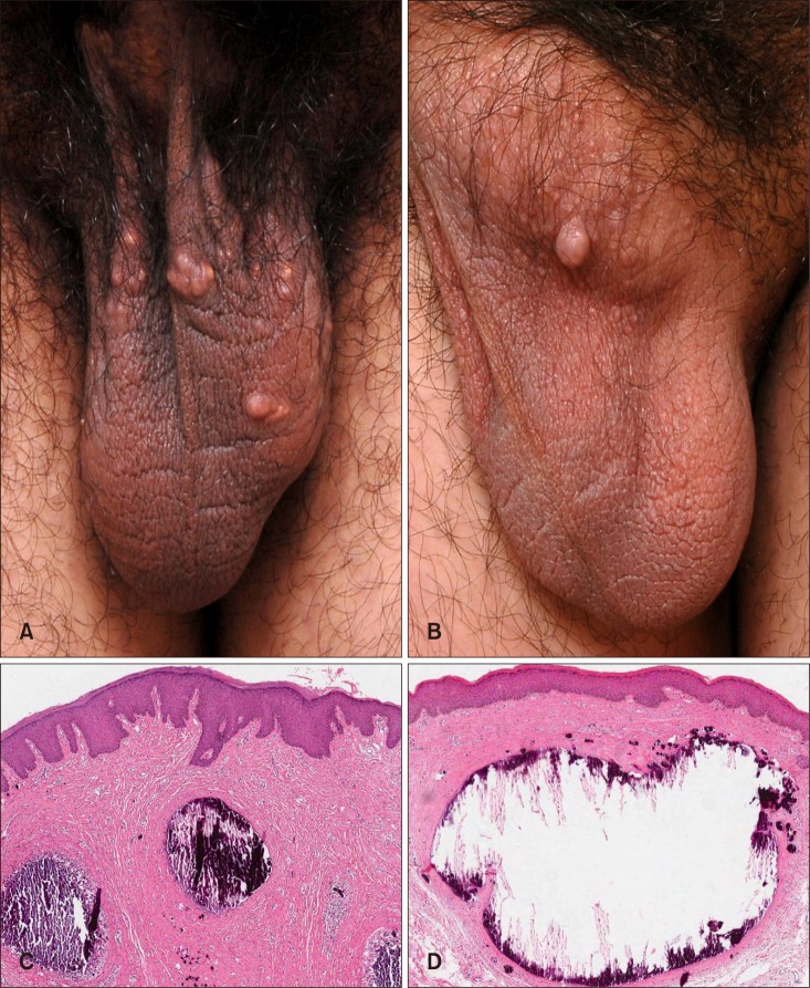
Fig. 1 (A, B) Several yellowish or whitish papules and nodules were present on the scrotums of both patients. (C, D) On skin biopsy, calcium deposits and basophilic globules of different sizes were observed in the dermis. The globules were surrounded by histiocytes and an inflammatory giant cell reaction was seen (H&E, ×40).
Reference
-
1. Shapiro L, Platt N, Torres-Rodríguez VM. Idiopathic calcinosis of the scrotum. Arch Dermatol. 1970; 102:199–204. PMID: 5464321.
Article2. Chiummariello S, Figus A, Menichini G, Bellezza G, Alfano C. Scrotal calcinosis: a very rare multiple clinical presentation. Clin Exp Dermatol. 2009; 34:e795–e797. PMID: 19817761.
Article3. Dubey S, Sharma R, Maheshwari V. Scrotal calcinosis: idiopathic or dystrophic? Dermatol Online J. 2010; 16:5.
Article
