Balloon dilation of congenital supravalvular pulmonic stenosis in a dog
- Affiliations
-
- 1Department of Clinical Sciences, College of Veterinary Medicine, Auburn University, 1220 Wire Road, Auburn, AL 36849, USA. szj0026@auburn.edu
- KMID: 2412596
- DOI: http://doi.org/10.4142/jvs.2017.18.1.111
Abstract
- Percutaneous balloon valvuloplasty is considered the standard of care for treatment of valvular pulmonic stenosis, a common congenital defect in dogs. Supravalvular pulmonic stenosis is a rare form of pulmonic stenosis in dogs and standard treatment has not been established. Although, there have been reports of successful treatment of supravalvular pulmonic stenosis with surgical and stenting techniques, there have been no reports of balloon dilation to treat dogs with this condition. Here, a case of supravalvular pulmonic stenosis diagnosed echocardiographically and angiographically in which a significant reduction in pressure gradient was achieved with balloon dilation alone is presented.
MeSH Terms
Figure
-
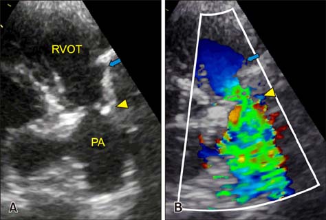
Fig. 1 Right parasternal short axis echocardiogram showing the right ventricular outflow tract (RVOT) and main pulmonary artery (PA). (A) The supravalvular lesion (yellow arrowhead) is distal to the pulmonary valve (blue arrow). (B) Color flow Doppler ultrasound shows turbulence originating at the level of the supravalvular stenosis.
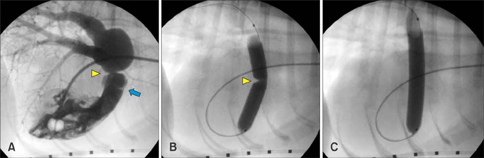
Fig. 2 Fluoroscopic images obtained before and during balloon dilation. (A) Right ventricular angiogram showing the pulmonary valve (blue arrow) and supravalvular pulmonic stenosis (yellow arrowhead). (B) Inflation of the balloon with diluted contrast solution across the stenotic lesion showing a discrete waist (yellow arrowhead) at the level of the supravalvular stenosis. (C) After full inflation of the balloon, complete loss of the waist is noted.
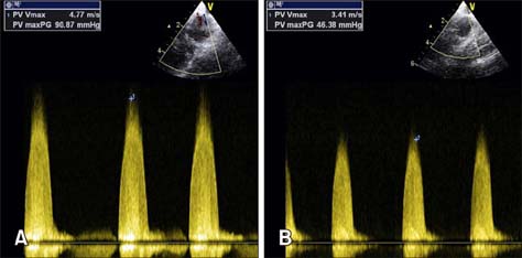
Fig. 3 Cranial transverse transesophageal echocardiogram of the right ventricular outflow tract. (A) Continuous wave Doppler across the right ventricular outflow tract prior to balloon dilation showing a peak pressure gradient of 90.87 mmHg. (B) Continuous wave Doppler across the right ventricular outflow tract after balloon dilation showing a decreased-peak pressure gradient of 46.38 mmHg.
Reference
-
1. Anderson M. What is your diagnosis? Right-sided cardiomegaly associated with supravalvular pulmonic stenosis. J Am Vet Med Assoc. 1992; 200:2013–2014.2. Bacha EA, Kalimi R, Starr JP, Quinones J, Koenig P. Autologous repair of supravalvar pulmonic stenosis. Ann Thorac Surg. 2004; 77:734–736.
Article3. Buchanan JW, Anderson JH, White RI. The 1st balloon valvuloplasty: an historical note. J Vet Intern Med. 2002; 16:116–117.
Article4. Dogan OF, Demircin M, Ozkutlu S, Pasaoglu I. Surgical management of infants with isolated supravalvular pulmonary stenosis: case reports. Heart Surg Forum. 2006; 9:E668–E674.
Article5. Estrada A, Moïse NS, Erb HN, McDonough SP, Renaud-Farrell S. Prospective evaluation of the balloon-to-annulus ratio for valvuloplasty in the treatment of pulmonic stenosis in the dog. J Vet Intern Med. 2006; 20:862–872.
Article6. Ford RB, Spaulding GL, Eyster GE. Use of an extracardiac conduit in the repair of supravalvular pulmonic stenosis in a dog. J Am Vet Med Assoc. 1978; 172:922–925.7. Griffiths LG, Bright JM, Chan KC. Transcatheter intravascular stent placement to relieve supravalvular pulmonic stenosis. J Vet Cardiol. 2006; 8:145–155.
Article8. Johnson MS, Martin M. Results of balloon valvuloplasty in 40 dogs with pulmonic stenosis. J Small Anim Pract. 2004; 45:148–153.
Article9. Lee ML, Chiu IS. Two-stage endovascular balloon angioplasty and stent implantation for supravalvular pulmonary stenosis in a 33-month-old boy incurring congestive heart failure following arterial switch operation. Acta Cardiol. 2013; 68:226–230.
Article10. Luciani A, Sconza S, Guglielmini C. Supravalvular PA stenosis (PAS) of probable congenital origin. J Am Vet Med Assoc. 2011; 239:1415–1416.11. McDevitt H, Stauthammer C, Leeder D, Hanson M, Olson J, Gruenstein D. Palliative balloon angioplasty in a cat with right pulmonary arterial branch stenoses and concurrent absence of the left pulmonary artery. J Vet Cardiol. 2013; 15:211–216.
Article12. Milo S, Fiegel A, Shem-Tov A, Neufeld HN, Goor DA. Hour-glass deformity of the pulmonary valve: a third type of pulmonary valve stenosis. Br Heart J. 1988; 60:128–133.
Article13. Rosales AM, Lock JE, Perry SB, Geggel RL. Interventional catheterization management of perioperative peripheral pulmonary stenosis: balloon angioplasty or endovascular stenting. Catheter Cardiovasc Interv. 2002; 56:272–277.
Article14. Saxena A, Fong LV, Ogilvie BC, Keeton BR. Use of balloon dilatation to treat supravalvar pulmonary stenosis developing after anatomical correction for complete transposition. Br Heart J. 1990; 64:151–155.
Article15. Soda A, Tanaka R, Saida Y, Yamane Y. Successful surgical correction of supravalvular pulmonary stenosis under beating heart using a cardiopulmonary bypass system in a dog. J Vet Med Sci. 2009; 71:203–206.
Article
- Full Text Links
-
- Actions
-
Cited
- CITED
-
- Close
- Share
-
- Similar articles
-
- A Case of Balloon Valvuloplasty using Three Balloon Catheters in a Child with Pulmonic Stenosis
- A Case of Supravalvular Aortic Stenosis
- A Case of Supravalvular and Valvular Aortic Stenosis
- Balloon valvuloplasty for severe subaortic stenosis in a Pomeranian dog
- Supravalvular Aortic Stenosis with Aortic Regurgitation
