Repetitive ultrasonographic assessment of adrenal size and shape changes: a clue for an asymptomatic sex hormone-secreting adenoma
- Affiliations
-
- 1Department of Veterinary Medical Imaging, College of Veterinary Medicine, Chonnam National University, Gwangju 61186, Korea. imsono@jnu.ac.kr
- 2Department of Veterinary Pathology, College of Veterinary Medicine, Chonnam National University, Gwangju 61186, Korea.
- 3Chonnam National University Veterinary Teaching Hospital, Gwangju 61186, Korea.
- KMID: 2412595
- DOI: http://doi.org/10.4142/jvs.2017.18.1.105
Abstract
- Diagnosis of an adrenal tumor without typical clinical signs related to hyperadrenocorticism and elevated alkaline phosphatase is challenging. This report describes a sex hormone-secreting adrenal tumor in a 10-year-old castrated male Shih Tzu evaluated through repetitive ultrasonographic examination. An adrenocorticotropic hormone stimulation test revealed elevated concentrations of androstenedione and 17-hydroxyprogesterone but a normal cortisol concentration. A mass was surgically excised and adenoma was diagnosed histopathologically. In the present case, adrenal tumor was strongly suspected based on a gradual increase in adrenal size and a change from peanut shape to an irregular mass on repetitive ultrasonography. Repetitive ultrasonographic examination of the adrenal gland is recommended when an abnormal ultrasonographic appearance of adrenal gland is identified, even in an asymptomatic dog.
MeSH Terms
Figure
-
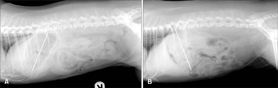
Fig. 1 Right lateral abdominal radiographs of the dog at first presentation (A) and approximately 20 months later (B). Note the increased liver size in (B) compared with that in (A). Liver size was determined based on the gastric axis (white line).
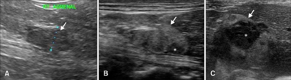
Fig. 2 Serial ultrasonography of the right adrenal gland at the first presentation (A), at 2 months (B), and approximately 20 months later (C). The size of the right adrenal gland (arrow) had increased and had changed to form a mass-like lesion with heterogeneous echotexture (*) and an irregular margin.
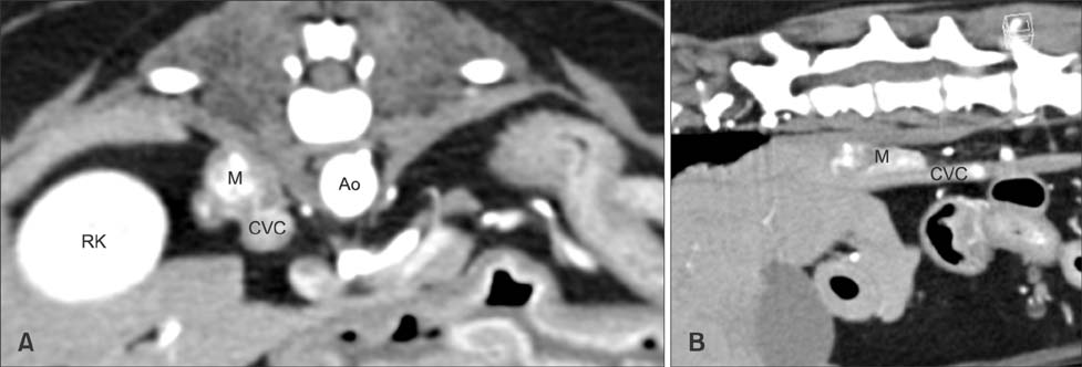
Fig. 3 Transverse image (A) and multiplanar reconstructed oblique sagittal plane image (B) from post-contrast abdominal computed tomography. Right adrenal mass was close to the wall of the caudal vena cava but did not invade it. Ao, aorta; RK, right kidney; CVC, caudal vena cava; M, mass.

Fig. 4 Representative image of histopathologic findings of the right adrenal tumor. Tumor cells were large and polyhedral with prominent nuclei and densely eosinophilic or had markedly vacuolated cytoplasm. H&E stain. Scale bar = 500 µm.
Reference
-
1. Besso JG, Penninck DG, Gliatto JM. Retrospective ultrasonographic evaluation of adrenal lesions in 26 dogs. Vet Radiol Ultrasound. 1997; 38:448–455.
Article2. Choi J, Kim H, Yoon J. Ultrasonographic adrenal gland measurements in clinically normal small breed dogs and comparison with pituitary-dependent hyperadrenocorticism. J Vet Med Sci. 2011; 73:985–989.
Article3. Davis MK, Schochet RA, Wrigley R. Ultrasonographic identification of vascular invasion by adrenal tumors in dogs. Vet Radiol Ultrasound. 2012; 53:442–445.
Article4. Feldman EC, Nelson RW, Feldman MS. Use of low- and high-dose dexamethasone tests for distinguishing pituitary-dependent from adrenal tumor hyperadrenocorticism in dogs. J Am Vet Med Assoc. 1996; 209:772–775.5. Gilor C, Graves TK. Interpretation of laboratory tests for canine Cushing's syndrome. Top Companion Anim Med. 2011; 26:98–108.
Article6. Gójska-Zygner O, Lechowski R, Zygner W. Functioning unilateral adrenocortical carcinoma in a dog. Can Vet J. 2012; 53:623–625.7. Grooters AM, Biller DS, Theisen SK, Miyabayashi T. Ultrasonographic characteristics of the adrenal glands in dogs with pituitary-dependent hyperadrenocorticism: comparison with normal dogs. J Vet Intern Med. 1996; 10:110–115.
Article8. Kyles AE, Feldman EC, De Cock HEV, Kass PH, Mathews KG, Hardie EM, Nelson RW, Ilkiw JE, Gregory CR. Surgical management of adrenal gland tumors with and without associated tumor thrombi in dogs: 40 cases (1994-2001). J Am Vet Med Assoc. 2003; 223:654–662.
Article9. Massari F, Nicoli S, Romanelli G, Buracco P, Zini E. Adrenalectomy in dogs with adrenal gland tumors: 52 cases (2002-2008). J Am Vet Med Assoc. 2011; 239:216–221.
Article10. Norman EJ, Thompson H, Mooney CT. Dynamic adrenal function testing in eight dogs with hyperadrenocorticism associated with adrenocortical neoplasia. Vet Rec. 1999; 144:551–554.
Article11. Schultz RM, Wisner ER, Johnson EG, MacLeod JS. Contrast-enhanced computed tomography as a preoperative indicator of vascular invasion from adrenal masses in dogs. Vet Radiol Ultrasound. 2009; 50:625–629.
Article12. Schwartz P, Kovak JR, Koprowski A, Ludwig LL, Monette S, Bergman PJ. Evaluation of prognostic factors in the surgical treatment of adrenal gland tumors in dogs: 41 cases (1999-2005). J Am Vet Med Assoc. 2008; 232:77–84.
Article13. Syme HM, Scott-Moncrieff JC, Treadwell NG, Thompson MF, Snyder PW, White MR, Oliver JW. Hyperadrenocorticism associated with excessive sex hormone production by an adrenocortical tumor in two dogs. J Am Vet Med Assoc. 2001; 219:1725–1728.
Article
- Full Text Links
-
- Actions
-
Cited
- CITED
-
- Close
- Share
-
- Similar articles
-
- MRI of the TSH(Thyroid Stimulating Hormone) -Secreting Pituitary Adenoma
- Microsurgical Transsphenoidal Approach for Pituitary Denomas
- Ultrasonographic measurement of the neonatal adrenal glands
- Transsphenoidal Microsurgery of Pituitary Adenoma
- A Case of Thyrotropin - Secreting Pituitary Adenoma with Normal alpha-subunit / TSH Molar Ratio
