Histopathologic findings in uteri and ovaries collected from clinically healthy dogs at elective ovariohysterectomy: a cross-sectional study
- Affiliations
-
- 1INCA-CES Research Group, School of Veterinary Medicine and Zootechny, CES University, Medellin 050021, Colombia.
- 2Veterinary Teaching Hospital, CES University, Envigado 0555427, Colombia.
- 3Research Group on Veterinary Sciences Centauro, School of Veterinary Medicine, Faculty of Agrarian Sciences, University of Antioquia, Medellin 050010, Colombia. juan.maldonado@udea.edu.co
- 4Laboratory of Veterinary Pathology, School of Veterinary Medicine, Faculty of Agrarian Sciences, University of Antioquia, Medellin 050010, Colombia.
- KMID: 2412459
- DOI: http://doi.org/10.4142/jvs.2017.18.3.407
Abstract
- Opinions on ovariohysterectomy (OHE) of bitches vary depending on region and country. In this descriptive, prospective cross-sectional study, uterine tracts and ovaries exhibiting gross pathologic findings (n = 76) were collected post-surgery from a reference population of 3,600 bitches (2.11% incidence) that underwent elective OHE during September to November 2013 and evaluated by histopathology examination. Data were evaluated by using descriptive statistics and chi-squared tests. Bitches were of crossbred background with average age 5 years (range 0.6-8.0 years) and most were nulliparous (69.7%) with no anamnesis of reproductive diseases (81.6%). Frequencies of proestrus, estrus, and diestrus were 42.1%, 6.6%, and 19.7%, respectively. The presence of mammary gland masses (5.3%) significantly correlated with histopathologic findings in ovaries and age of the bitch (p < 0.05). Predominant uterine histopathologies included cystic endometrial hyperplasia, periglandular fibrosis, lymphoplasmocytary endometritis, and adenomyosis (19.7%, 14.5%, 4.0%, and 2.6%, respectively). In ovaries, hyperplasia of rete ovarii, follicular cysts, oophoritis, adenoma of the rete ovarii, cysts of superficial structures, and granulosa cell tumors (10.5%, 10.5%, 7.9%, 4.0%, 2.6%, and 2.6%, respectively) were observed. The results reveal the presence of subclinical pathologies in healthy bitches, suggesting that OHE at an early age is beneficial for prevention of reproductive pathologies.
Keyword
MeSH Terms
-
Animals
Cross-Sectional Studies
Dog Diseases/pathology
Dogs
Endometrial Hyperplasia/pathology/veterinary
Endometritis/pathology/veterinary
Female
Hysterectomy/methods/*veterinary
Ovarian Diseases/pathology/veterinary
Ovarian Neoplasms/pathology/veterinary
Ovariectomy/methods/*veterinary
Ovary/*pathology/surgery
Uterus/*pathology/surgery
Figure
-
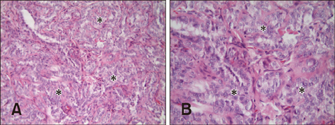
Fig. 1 Proliferative disorders found in ovaries. (A) Epithelial proliferation of the rete ovarii (asterisks). (B) Stratification of the rete ovarii showing epithelial proliferation in a different ovary from that of panel A. H&E stain. 200× (A), 400× (B).
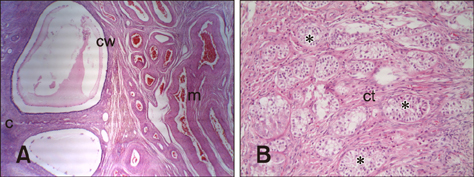
Fig. 2 Follicle cysts. (A) Microscopic aspect of the cyst wall (cw), ovary cortex (c), ovary medulla (m). (B) Granulosa cell tumor exhibiting a sertoli pattern; connective tissue (ct). H&E stain. 40× (A), 200× (B).
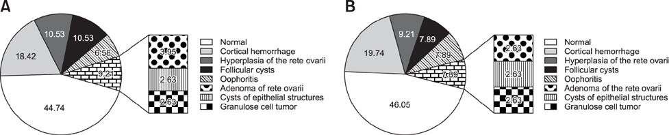
Fig. 3 Frequency of occurrence of microscopic lesions in the left (A) and right (B) ovaries at elective ovariohysterectomy in clinically healthy bitches with uteri containing gross pathologic findings.

Fig. 4 Cystic endometrial hyperplasia. (A and B) Dilated cystic glands (asterisk) from two different cases. Endometrium (em). Endometrium fibrosis. (C and D) severe periglandular fibrosis (asterisks) with abundant active fibroblasts (arrow). Endometrium (em) and endometrial glands (g). H&E stain. 40× (A, B, and D), 200× (C).
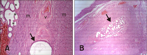
Fig. 5 (A) Adenomyosis showing cystic glands (arrow); myometrium (m), vascular layer (v) and hyperplasic endometrial glands with cysts (asterisks). (B) Adenomyosis, in which groups of endometrial glands appears embedded into the myometrium (arrow). H&E stain. 100× (A), 40× (B).
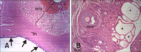
Fig. 6 (A) Mesonephric duct cyst showing the endometrium (em), myometrium (m) and cyst wall (arrows). (B) Cysts of superficial epithelium containing cystic structures of several sizes are seen in the ovarian cortex (asterisks). Cumulusoocyte-complex (coc) are present. H&E stain. 400× (A), 100× (B).
Reference
-
1. Arora N, Sandford J, Browning GF, Sandy JR, Wright PJ. A model for cystic endometrial hyperplasia/pyometra complex in the bitch. Theriogenology. 2006; 66:1530–1536.
Article2. Balka G, Szabó L, Jakab C. First report of an endometrial adenoacanthoma in a dog. Acta Vet Hung. 2011; 59:225–236.
Article3. Bhatti SF, Rao NA, Okkens AC, Mol JA, Duchateau L, Ducatelle R, van den Ingh TS, Tshamala M, Van Ham LM, Coryn M, Rijnberk A, Kooistra HS. Role of progestin-induced mammary-derived growth hormone in the pathogenesis of cystic endometrial hyperplasia in the bitch. Domest Anim Endocrinol. 2007; 33:294–312.
Article4. Bostedt H, Jung C, Wehrend A, Boryzcko Z. [Clinical and endocrinological findings of bitches with ovarian cyst syndrome]. Schweiz Arch Tierheilkd. 2013; 155:543–550. German.5. Brønden LB, Nielsen SS, Toft N, Kristensen AT. Data from the Danish veterinary cancer registry on the occurrence and distribution of neoplasms in dogs in Denmark. Vet Rec. 2010; 166:586–590.
Article6. Colimon KM. [Fundamentals of Epidemiology]. 3a ed. Medellín: Corporación para Investigaciones Biológicas;2010. p. 551. Spanish.7. Concannon PW, Spraker TR, Casey HW, Hansel W. Gross and histopathologic effects of medroxyprogesterone acetate and progesterone on the mammary glands of adult beagle bitches. Fertil Steril. 1981; 36:373–387.
Article8. De Bosscher H, Ducatelle R, Tshamala M, Coryn M. Changes in sex hormone receptors during administration of progesterone to prevent estrus in the bitch. Theriogenology. 2002; 58:1209–1217.
Article9. De Bosschere H, Ducatelle R, Vermeirsch H, Van Den Broeck W, Coryn M. Cystic endometrial hyperplasia-pyometra complex in the bitch: should the two entities be disconnected. Theriogenology. 2001; 55:1509–1519.
Article10. Ferreira de la Cuesta G. [Veterinary Pathology]. lst ed. Medellin: Editorial Universidad de Antioquia;2003. p. 526–530. Spanish.11. Gómez B, Ramírez M, Maldonado-Estrada J. Presence of lung metastases in bitches affected by malignant mammary neoplasms in Medellin (Colombia). Rev MVZ Cordoba. 2012; 17:2983–2990.12. González-Domínguez MS, Fernández LB, Saldarriaga S, Aranzazu-Taborda D, Maldonado-Estrada JG. [Infertility in a bitch with history of recurrent reproductive failure associated with granulosa cell tumor]. Rev Colomb Cienc Pecu. 2005; 18:258–268. Spanish.13. Groppetti D, Pecile A, Arrighi S, Di Giancamillo A, Cremonesi F. Endometrial cytology and computerized morphometric analysis of epithelial nuclei: a useful tool for reproductive diagnosis in the bitch. Theriogenology. 2010; 73:927–941.
Article14. Günzel-Apel AR, Buschhaus J, Urhausen C, Masal C, Wolf K, Meyer-Lindenberg A, Piechotta M, Beyerbach M, Schoon HA. [Clinical signs, diagnostic approach and therapy for the so-called ovarian remnant syndrome in the bitch]. Tierarztl Prax Ausg K Kleintiere Heimtiere. 2012; 40:35–42. German.15. Hagman R, Lagerstedt AS, Hedhammar Å, Egenvall A. A breed-matched case-control study of potential risk factors for canine pyometra. Theriogenology. 2011; 75:1251–1257.
Article16. Ichimura R, Shibutani M, Mizukami S, Suzuki T, Shimada Y, Mitsumori K. A case report of an uncommon sex-cord stromal tumor consisted of luteal and sertoli cells in a spayed bitch. J Vet Med Sci. 2010; 72:229–234.
Article17. Knauf Y, Bostedt H, Failing K, Knauf S, Wehrend A. Gross pathology and endocrinology of ovarian cysts in bitches. Reprod Domest Anim. 2014; 49:463–468.
Article18. Kustritz MV. Determining the optimal age for gonadectomy of dogs and cats. J Am Vet Med Assoc. 2007; 231:1665–1675.
Article19. Marino G, Barna A, Rizzo S, Zanghì A, Catone G. Endometrial polyps in the bitch: a retrospective study of 21 cases. J Comp Pathol. 2013; 149:410–416.
Article20. McEntee MC. Reproductive oncology. Clin Tech Small Anim Pract. 2002; 17:133–149.
Article21. McGavin MD, Zachary JF. Pathologic Basis of Veterinary Disease. 5th ed. St. Louis: Elsevier;2012. p. 1079–1092.22. McKay SA, Farnworth MJ, Waran NK. Current attitudes toward, and incidence of, sterilization of cats and dogs by caregivers (owners) in Auckland, New Zealand. J Appl Anim Welf Sci. 2009; 12:331–344.
Article23. Merlo DF, Rossi L, Pellegrino C, Ceppi M, Cardellino U, Capurro C, Ratto A, Sambucco PL, Sestito V, Tanara G, Bocchini V. Cancer incidence in pet dogs: findings of the Animal Tumor Registry of Genoa, Italy. J Vet Intern Med. 2008; 22:976–984.
Article24. Meuten DJ, editor. Tumors in Domestic Animals. 4th ed. Ames: Iowa State Press;2003. p. 176–179.25. Mir F, Fontaine E, Albaric O, Greer M, Vannier F, Schlafer DH, Fontbonne A. Findings in uterine biopsies obtained by laparotomy from bitches with unexplained infertility or pregnancy loss: an observational study. Theriogenology. 2013; 79:312–322.
Article26. Misdorp W. Progestagens and mammary tumours in dogs and cats. Acta Endocrinol (Copenh). 1991; 125:Suppl 1. 27–31.27. Nickel RF, Okkens AC, van der Gaag I, van Haaften B. Oophoritis in a dog with abnormal corpus luteum function. Vet Rec. 1991; 128:333–334.
Article28. Niskanen M, Thrusfield MV. Associations between age, parity, hormonal therapy and breed, and pyometra in Finnish dogs. Vet Rec. 1998; 143:493–498.
Article29. Ortega-Pacheco A, Gutiérrez-Blanco E, Jiménez-Coello M. Common lesions in the female reproductive tract of dogs and cats. Vet Clin North Am Small Anim Pract. 2012; 42:547–559.
Article30. Patnaik AK, Greenlee PG. Canine ovarian neoplasms: a clinicopathologic study of 71 cases, including histology of 12 granulosa cell tumors. Vet Pathol. 1987; 24:509–514.
Article31. Patsikas M, Papazoglou LG, Jakovljevic S, Papaioannou NG, Papadopoulou PL, Soultani CB, Chryssogonidis IA, Kouskouras KA, Tziris NE, Charitanti AA. Radiographic and ultrasonographic findings of uterine neoplasms in nine dogs. J Am Anim Hosp Assoc. 2014; 50:330–337.
Article32. Pena FJ, Gines JA, Duque J, Vieitez V, Martinez-Pérez R, Madejón L, Nuñez Martinez I, Moran JM, Fernández-García S. Endometrial adenocarcinoma and mucometra in a 6-year-old Alaska Malamute dog. Reprod Domest Anim. 2006; 41:189–190.
Article33. Pires MA, Seixas F, Palmeira C, Payan-Carreira R. Histopathologic and immunohistochemical exam in one case of canine endometrial adenocarcinoma. Reprod Domest Anim. 2010; 45:545–549.
Article34. Pretzer SD. Clinical presentation of canine pyometra and mucometra: a review. Theriogenology. 2008; 70:359–363.
Article35. Schneider R, Dorn CR, Taylor DON. Factors influencing canine mammary cancer development and postsurgical survival. J Natl Cancer Inst. 1969; 43:1249–1261.36. Sleeckx N, de Rooster H, Veldhuis Kroeze EJB, Van Ginneken C, Van Brantegem L. Canine mammary tumors, an overview. Reprod Domest Anim. 2011; 46:1112–1131.37. Smith FO. Canine pyometra. Theriogenology. 2006; 66:610–612.
Article38. Van Goethem B, Schaefers-Okkens A, Kirpensteijn J. Making a rational choice between ovariectomy and ovariohysterectomy in the dog: a discussion of the benefits of either technique. Vet Surg. 2006; 35:136–143.
Article39. Zanghì A, Catone G, Marino G, Quartuccio M, Nicòtina PA. Endometrial polypoid adenomyomatosis in a bitch with ovarian granulosa cell tumor and pyometra. J Comp Pathol. 2007; 136:83–86.
Article
- Full Text Links
-
- Actions
-
Cited
- CITED
-
- Close
- Share
-
- Similar articles
-
- Attenuated total reflection Fourier transform infrared as a primary screening method for cancer in canine serum
- Sterile Pyometra in Two Dogs
- A case of transient diabetes mellitus in a dog managed by ovariohysterectomy
- Evaluation of circulating IGF-I and IGFBP-3 as biomarkers for tumors in dogs
- Evaluation of circulating PD-1 and PD-L1 as diagnostic biomarkers in dogs with tumors
