Fixed Lunate Flexion Deformity in Distal Radius Fractures
- Affiliations
-
- 1Department of Orthopedic Surgery, Inje University Sanggye Paik Hospital, Inje University College of Medicine, Seoul, Korea.
- 2Department of Orthopedic Surgery, National Medical Center, Seoul, Korea. mdjsh@nate.com
- KMID: 2412352
- DOI: http://doi.org/10.4055/cios.2016.8.2.228
Abstract
- Carpal malalignments in malunion of distal radius fracture are considered as an adaptive response of the carpus to loss of normal architecture of the distal radius. This condition leads to mechanical overload, ligament attenuation and progressive dynamic instability around the wrist joint. Radial corrective osteotomy is suggested as a treatment option of carpal malalignment after distal radius malunion. In radiocarpal malalignment, the lunate is usually observed in flexion in contrast to its extension posture in the more common midcarpal malalignment. We report two cases of fixed lunate flexion deformity after a distal radius fracture, in which reduction and fixation of fresh fracture or corrective osteotomy of malunion were not successful. Arthritic changes were observed in the radiolunate joint on arthroscopy. Thus, fixed flexion deformity of the lunate might be associated with posttraumatic arthritic change in the radiolunate joint.
MeSH Terms
Figure
-
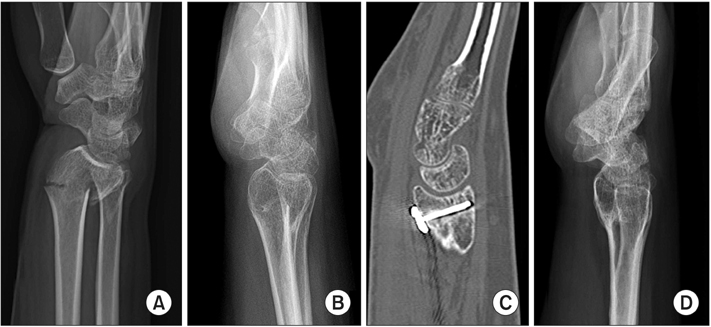
Fig. 1 Initial (A), preoperative (B), and postoperative (C, D) radiographs of case 1 show the preoperative dorsal tilt of 27° (B), the postoperative volar tilt of 3° (C, D), and the flexion of the lunate after corrective osteotomy of the distal radius.

Fig. 2 Wrist arthroscopy of case 1 shows articular debris and fibrillation of distal radius (A) and cartilage denudation of the lunate (B).
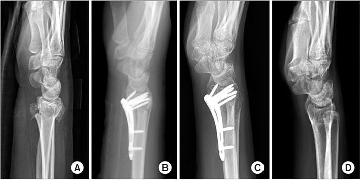
Fig. 3 Preoperative (A) and immediate postoperative (B) radiographs of case 2 show normal alignment of the lunate. However, flexion of the lunate was evident at 6 weeks postoperatively (C) and persisted at the last follow-up (D).
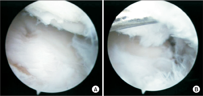
Fig. 4 Arthroscopy of case 2 shows wear and irregularities of the articular surface of the radius (A) and the lunate (B).
Reference
-
1. Gupta A, Batra S, Jain P, Sharma SK. Carpal alignment in distal radial fractures. BMC Musculoskelet Disord. 2002; 3:14.
Article2. Verhaegen F, Degreef I, De Smet L. Evaluation of corrective osteotomy of the malunited distal radius on midcarpal and radiocarpal malalignment. J Hand Surg Am. 2010; 35(1):57–61.
Article3. De Smet L, Verhaegen F, Degreef I. Carpal malalignment in malunion of the distal radius and the effect of corrective osteotomy. J Wrist Surg. 2014; 3(3):166–170.4. Sennwald G, Fischer W, Stahelin A. Malunion of the distal radius and its treatment: apropos of 122 radii. Int Orthop. 1992; 16(1):45–51.5. Park MJ, Cooney WP 3rd, Hahn ME, Looi KP, An KN. The effects of dorsally angulated distal radius fractures on carpal kinematics. J Hand Surg Am. 2002; 27(2):223–232.
Article6. Bushnell BD, Bynum DK. Malunion of the distal radius. J Am Acad Orthop Surg. 2007; 15(1):27–40.
Article7. McQueen MM, Wakefield A. Distal radial osteotomy for malunion using non-bridging external fixation: good results in 23 patients. Acta Orthop. 2008; 79(3):390–395.
Article8. Batra S, Debnath U, Kanvinde R. Can carpal malalignment predict early and late instability in nonoperatively managed distal radius fractures. Int Orthop. 2008; 32(5):685–691.
Article
- Full Text Links
-
- Actions
-
Cited
- CITED
-
- Close
- Share
-
- Similar articles
-
- Does the Morphological Type of the Lunate Affect Surgical Outcomes in Patients with Distal Radius Fractures?
- Complications of Distal Radius Fracture
- Volar Plating of Distal Radius Fractures
- Salter-Harris Type IV Physeal Fracture of the Distal Radius: A Case Report
- Arthroscopically Assisted Reduction of Distal Radius Fractures
