Clin Orthop Surg.
2016 Jun;8(2):175-180. 10.4055/cios.2016.8.2.175.
The Role of Lunate Morphology on Scapholunate Instability and Fracture Location in Patients Treated for Scaphoid Nonunion
- Affiliations
-
- 1Department of Orthopaedic Surgery, Chonnam National University Medical School, Gwangju, Korea. mskim@jnu.ac.kr
- 2Department of Orthopaedics, Yale University Medical School, New Haven, CT, USA.
- KMID: 2412343
- DOI: http://doi.org/10.4055/cios.2016.8.2.175
Abstract
- BACKGROUND
To determine the association between lunate morphology and the scapholunate instability using radiographic images, and investigate the association between lunate morphology and scaphoid fracture location.
METHODS
Between January 2003 and December 2011, we retrospectively evaluated the plain radiographs and computed tomography (CT) images of 70 patients who underwent surgical intervention for a scaphoid nonunion, in order to determine the association between lunate type (I or II) and scapholunate instability or scaphoid fracture location. We determined the scaphoid fracture location using the fragment ratio and measured the radiolunate angle and capitate-triquetrum (C-T) distance.
RESULTS
A type II lunate was present in 68.6% (48 of 70 cases). Mean fragment ratio of fracture location was 50.6% in the type II lunate group and 56.2% in the type I lunate group (p = 0.032). Sixteen of the 70 patients had dorsal intercalated segmental instability (DISI) deformities. Nine of 22 cases showed DISI deformity in type I lunate and 7 of 48 cases showed DISI deformity in type II lunate (p = 0.029). However, there were no significant differences between the presence of DISI deformity and fracture location (p = 0.15). Morphologic comparisons by both plain radiography and CT indicated a mean C-T distance in the type I lunate group (22 cases) of 2.3 mm and 5.0 mm in the type II lunate group (48 cases). The C-T distances were significantly correlated with lunate morphology (p = 0.001).
CONCLUSIONS
A type II lunate was associated with low incidence of DISI deformity and proximal location of fracture in patients presenting with a scaphoid nonunion.
MeSH Terms
-
Adolescent
Adult
Female
*Fractures, Bone/diagnostic imaging/physiopathology
Humans
Joint Instability/diagnostic imaging/physiopathology
*Lunate Bone/anatomy & histology/diagnostic imaging/physiopathology
Male
Middle Aged
Retrospective Studies
*Scaphoid Bone/diagnostic imaging/injuries/physiopathology
Tomography, X-Ray Computed
*Wrist Injuries/diagnostic imaging/physiopathology
Young Adult
Figure
-

Fig. 1 Type I lunate with a single midcarpal articulating facet with the capitate (A, B) and type II lunate with two midcarpal articulating facets, one with the capitate and the other with the hamate (C, D).
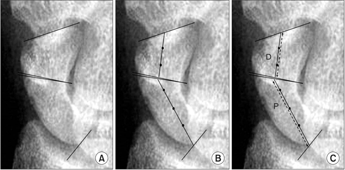
Fig. 2 (A) Fragment ratio. Horizontal lines were drawn to define the extent of the fragment. (B) The middle of the fragment was identified and a midline was drawn connecting the two horizontal lines. (C) The lengths of these lines (P: proximal fragment, D: distal fragment) were measured to determine the fragment size. Fragment ratio = P / (P + D).
Reference
-
1. Larsen CF, Amadio PC, Gilula LA, Hodge JC. Analysis of carpal instability: I. description of the scheme. J Hand Surg Am. 1995; 20(5):757–764.
Article2. Sennwald G, Segmuller G. Anatomic basis of a new concept of stability of the carpus. Int Orthop. 1986; 10(1):25–30.3. Haase SC, Berger RA, Shin AY. Association between lunate morphology and carpal collapse patterns in scaphoid nonunions. J Hand Surg Am. 2007; 32(7):1009–1012.
Article4. Viegas SF. The lunatohamate articulation of the midcarpal joint. Arthroscopy. 1990; 6(1):5–10.
Article5. Viegas SF. Variations in the skeletal morphologic features of the wrist. Clin Orthop Relat Res. 2001; (383):21–31.
Article6. Viegas SF, Patterson RM, Hokanson JA, Davis J. Wrist anatomy: incidence, distribution, and correlation of anatomic variations, tears, and arthrosis. J Hand Surg Am. 1993; 18(3):463–475.
Article7. Viegas SF, Patterson RM, Todd PD, McCarty P. Load mechanics of the midcarpal joint. J Hand Surg Am. 1993; 18(1):14–18.
Article8. Viegas SF. Advances in the skeletal anatomy of the wrist. Hand Clin. 2001; 17(1):1–11.
Article9. Nakamura K, Beppu M, Patterson RM, Hanson CA, Hume PJ, Viegas SF. Motion analysis in two dimensions of radial-ulnar deviation of type I versus type II lunates. J Hand Surg Am. 2000; 25(5):877–888.
Article10. Galley I, Bain GI, McLean JM. Influence of lunate type on scaphoid kinematics. J Hand Surg Am. 2007; 32(6):842–847.
Article11. Nakamura K, Patterson RM, Moritomo H, Viegas SF. Type I versus type II lunates: ligament anatomy and presence of arthrosis. J Hand Surg Am. 2001; 26(3):428–436.
Article12. Sagerman SD, Hauck RM, Palmer AK. Lunate morphology: can it be predicted with routine x-ray films? J Hand Surg Am. 1995; 20(1):38–41.
Article13. Pfirrmann CW, Theumann NH, Chung CB, Trudell DJ, Resnick D. The hamatolunate facet: characterization and association with cartilage lesions—magnetic resonance arthrography and anatomic correlation in cadaveric wrists. Skeletal Radiol. 2002; 31(8):451–456.
Article14. Viegas SF, Wagner K, Patterson R, Peterson P. Medial (hamate) facet of the lunate. J Hand Surg Am. 1990; 15(4):564–571.
Article15. Gilula LA, Weeks PM. Post-traumatic ligamentous instabilities of the wrist. Radiology. 1978; 129(3):641–651.
Article16. Nakamura R, Hori M, Imamura T, Horii E, Miura T. Method for measurement and evaluation of carpal bone angles. J Hand Surg Am. 1989; 14(2 Pt 2):412–416.
Article17. Compson JP, Waterman JK, Heatley FW. The radiological anatomy of the scaphoid. Part 2: Radiology. J Hand Surg Br. 1997; 22(1):8–15.18. Ramamurthy C, Cutler L, Nuttall D, Simison AJ, Trail IA, Stanley JK. The factors affecting outcome after non-vascular bone grafting and internal fixation for nonunion of the scaphoid. J Bone Joint Surg Br. 2007; 89(5):627–632.
Article19. Nakamura K, Beppu M, Matsushita K, Arai T, Ide T. Biomechanical analysis of the stress force on midcarpal joint in Kienbock's disease. Hand Surg. 1997; 2(2):101–115.20. McLean JM, Bain GI, Watts AC, Mooney LT, Turner PC, Moss M. Imaging recognition of morphological variants at the midcarpal joint. J Hand Surg Am. 2009; 34(6):1044–1055.
Article21. Burgess RC. Anatomic variations of the midcarpal joint. J Hand Surg Am. 1990; 15(1):129–131.
Article22. Malik AM, Schweitzer ME, Culp RW, Osterman LA, Manton G. MR imaging of the type II lunate bone: frequency, extent, and associated findings. AJR Am J Roentgenol. 1999; 173(2):335–338.
Article23. Aufauvre B, Herzberg G, Garret J, Berthonneaud E, Dimnet J. A new radiographic method for evaluation of the position of the carpus in the coronal plane: results in normal subjects. Surg Radiol Anat. 1999; 21(6):383–385.
Article24. Bain GI, Clitherow HD, Millar S, et al. The effect of lunate morphology on the 3-dimensional kinematics of the carpus. J Hand Surg Am. 2015; 40(1):81–89.e1.
Article25. Moritomo H, Murase T, Oka K, Tanaka H, Yoshikawa H, Sugamoto K. Relationship between the fracture location and the kinematic pattern in scaphoid nonunion. J Hand Surg Am. 2008; 33(9):1459–1468.
Article
- Full Text Links
-
- Actions
-
Cited
- CITED
-
- Close
- Share
-
- Similar articles
-
- Radiological Analysis of the Scaphoid Shift Test
- Scapholunate Dissociation Associated with Intra-articular Fractures of Distal Radius
- Risk Factors of Nonunion after Surgical Fixation of Acute Scaphoid Fractures
- Anterior Interpositional Grafting for Scaphoid Nonunion with Dorsal Instability Rattern
- Perilunate Injury Combined with Lunate Fracture
