Analysis of Sacrococcygeal Morphology in Koreans Using Computed Tomography
- Affiliations
-
- 1Department of Orthopedic Surgery and Traumatology, Cheju Halla General Hospital, Jeju, Korea. mingy9879@gmail.com
- KMID: 2412324
- DOI: http://doi.org/10.4055/cios.2016.8.4.412
Abstract
- BACKGROUND
The sacrococcygeal morphology of Arabs and Europeans has been studied using computed tomography (CT) or magnetic resonance imaging to determine the cause of coccydynia. Studies have suggested differences in sacrococcygeal morphology among ethnic groups. However, there are no data on the sacrococcygeal anatomy of Koreans.
METHODS
We conducted a retrospective analysis of 606 pelvic CT scans that were taken at Cheju Halla General Hospital between 2008 and 2014. Fractures of the sacrum or coccyx were excluded. Differences in the sacrococcygeal morphology among age groups stratified by decade of life and between genders were analyzed using sagittal plane pelvic CT scans. The morphological parameters studied were the sacral and coccygeal curved indexes, sacrococcygeal angle, intercoccygeal angle, coccygeal type, coccygeal segmental number, and sacrococcygeal fusion.
RESULTS
The average sacral and coccygeal curved indexes were 6.15 and 7.41, respectively. The average sacrococcygeal and intercoccygeal angles were 110° and 49°, respectively. Type II coccyx was most common, and the rate of sacrococcygeal fusion was 34%. There was a moderate positive correlation between age and the sacral curved index (r = 0.493, p = 0.000) and a weak negative correlation between age and the coccyx curved index (r = −0.257, p = 0.000). There was a weak negative correlation between age and the intercoccygeal angle (r = −0.187, p = 0.000). The average intercoccygeal angle in males and females was 53.9° and 44.7°, respectively.
CONCLUSIONS
The sacrum tended to be more curved and the coccyx straighter with age. The coccyx was straighter in females than males. Knowledge of the sacrococcygeal anatomy of Koreans will promote better understanding of anatomical differences among ethnicities and future studies on coccydynia.
Keyword
MeSH Terms
Figure
-

Fig. 1 Computed tomography images showing morphology of the sacrococcygeal region. (A) The coccygeal type is classified according to the angle between a line from the center of the first coccygeal segment to the tip of the last coccygeal segment and a vertical line. (B) Sacrococcygeal fusion (circle) is noted.
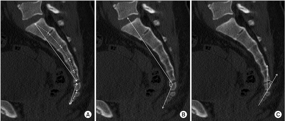
Fig. 2 Computed tomography images for morphometry of the sacrococcygeal region. (A) Straight (solid line) and curved (dotted line) lengths of each sacrum and coccyx. (B) Sacrococcygeal angle. (C) Intercoccygeal angle.
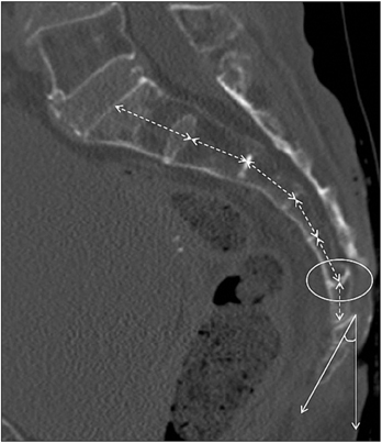
Fig. 3 In a 92-year-old female patient, the sacrum is curved (dotted line) and the coccyx is straight. Type I coccyx (angle) and sacrococcygeal fusion (circle) are noted.
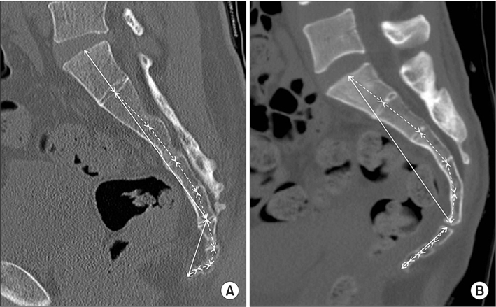
Fig. 4 Differences in sacrococcygeal anatomy between a male (A) and a female (B). In the female, the sacrum is more curved and the coccyx is straighter than in the male.
Reference
-
1. Maigne JY, Doursounian L, Chatellier G. Causes and mechanisms of common coccydynia: role of body mass index and coccygeal trauma. Spine (Phila Pa 1976). 2000; 25(23):3072–3079.2. Lirette LS, Chaiban G, Tolba R, Eissa H. Coccydynia: an overview of the anatomy, etiology, and treatment of coccyx pain. Ochsner J. 2014; 14(1):84–87.3. Patijn J, Janssen M, Hayek S, Mekhail N, Van Zundert J, van Kleef M. 14. Coccygodynia. Pain Pract. 2010; 10(6):554–559.4. Fogel GR, Cunningham PY 3rd, Esses SI. Coccygodynia: evaluation and management. J Am Acad Orthop Surg. 2004; 12(1):49–54.
Article5. Postacchini F, Massobrio M. Idiopathic coccygodynia: analysis of fifty-one operative cases and a radiographic study of the normal coccyx. J Bone Joint Surg Am. 1983; 65(8):1116–1124.
Article6. Maigne JY, Pigeau I, Roger B. Magnetic resonance imaging findings in the painful adult coccyx. Eur Spine J. 2012; 21(10):2097–2104.
Article7. Kim NH, Suk KS. Clinical and radiological differences between traumatic and idiopathic coccygodynia. Yonsei Med J. 1999; 40(3):215–220.
Article8. Nathan ST, Fisher BE, Roberts CS. Coccydynia: a review of pathoanatomy, aetiology, treatment and outcome. J Bone Joint Surg Br. 2010; 92(12):1622–1627.9. Woon JT, Stringer MD. Clinical anatomy of the coccyx: a systematic review. Clin Anat. 2012; 25(2):158–167.
Article10. Karadimas EJ, Trypsiannis G, Giannoudis PV. Surgical treatment of coccygodynia: an analytic review of the literature. Eur Spine J. 2011; 20(5):698–705.
Article11. Maigne JY, Lagauche D, Doursounian L. Instability of the coccyx in coccydynia. J Bone Joint Surg Br. 2000; 82(7):1038–1041.
Article12. Mouhsine E, Garofalo R, Chevalley F, et al. Posttraumatic coccygeal instability. Spine J. 2006; 6(5):544–549.
Article13. Marwan YA, Al-Saeed OM, Esmaeel AA, Kombar OR, Bendary AM, Azeem ME. Computed tomography-based morphologic and morphometric features of the coccyx among Arab adults. Spine (Phila Pa 1976). 2014; 39(20):E1210–E1219.
Article14. Woon JT, Maigne JY, Perumal V, Stringer MD. Magnetic resonance imaging morphology and morphometry of the coccyx in coccydynia. Spine (Phila Pa 1976). 2013; 38(23):E1437–E1445.
Article15. Woon JT, Perumal V, Maigne JY, Stringer MD. CT morphology and morphometry of the normal adult coccyx. Eur Spine J. 2013; 22(4):863–870.
Article16. Maigne JY, Tamalet B. Standardized radiologic protocol for the study of common coccygodynia and characteristics of the lesions observed in the sitting position: clinical elements differentiating luxation, hypermobility, and normal mobility. Spine (Phila Pa 1976). 1996; 21(22):2588–2593.
Article17. Przybylski P, Pankowicz M, Bockowska A, et al. Evaluation of coccygeal bone variability, intercoccygeal and lumbosacral angles in asymptomatic patients in multislice computed tomography. Anat Sci Int. 2013; 88(4):204–211.
Article18. Tague RG. Fusion of coccyx to sacrum in humans: prevalence, correlates, and effect on pelvic size, with obstetrical and evolutionary implications. Am J Phys Anthropol. 2011; 145(3):426–437.
Article19. Moore KL. Clinically oriented anatomy. 2nd ed. Baltimore, MD: Williams & Wilkins;1985.20. Gray H, Williams PL, Warwick R. Gray's anatomy. 36th ed. Edinburgh: Churchill-Livingstone;1980.21. Breathnach AS. Frazer's anatomy of the human skeleton. 6th ed. London: J & A Churchill;1965.22. Saluja PG. The incidence of ossification of the sacrococcygeal joint. J Anat. 1988; 156:11–15.23. Kerimoglu U, Dagoglu MG, Ergen FB. Intercoccygeal angle and type of coccyx in asymptomatic patients. Surg Radiol Anat. 2007; 29(8):683–687.
Article24. Balain B, Eisenstein SM, Alo GO, et al. Coccygectomy for coccydynia: case series and review of literature. Spine (Phila Pa 1976). 2006; 31(13):E414–E420.
Article
- Full Text Links
-
- Actions
-
Cited
- CITED
-
- Close
- Share
-
- Similar articles
-
- Coccygeal Morphology on Multislice Computed Tomography in a Tertiary Hospital in India
- Cystic Teratoma of the Sacrococcygeal Region in Adult: A Case Report
- Temporomandibular joint morphology in Korean using cone-beam computed tomography: influence of age and gender
- A Case of Sacrococcygeal Teratoma
- Sacrococcygeal Chordoma
