A Neglected Markedly Displaced Medial Epicondyle Fracture with Simultaneous Ulnar Nerve Palsy in an Adolescent
- Affiliations
-
- 1Division of Pediatric Orthopedics, Department of Orthopedic Surgery, Ain-Shams University Faculty of Medicine, Cairo, Egypt. tamer.ahmed@med.asu.edu.eg
- 2Department of Radiodiagnosis, Ain-Shams University Faculty of Medicine, Cairo, Egypt.
- KMID: 2412258
- DOI: http://doi.org/10.4055/cios.2017.9.4.542
Abstract
- Humeral medial epicondyle fractures constitute around 15% of pediatric elbow fractures. Up to 60% occur in association with elbow dislocations. Knowledge of potential imaging pitfalls when examining acute elbow fractures in children contributes significantly to accurate diagnosis. Nevertheless, management of missed pediatric medial epicondyle fractures has rarely been reported. We present an 11-year-old boy with a neglected and severely displaced medial epicondyle fracture with concurrent ulnar nerve palsy. We performed neural decompression, fragment excision, and muscular and capsuloligamentous reconstruction of the medial elbow. This study demonstrates that the surgical outcome of a late presenting fracture can be satisfactory in terms of function and neural recovery. It also underscores the importance of careful interpretation of elbow imaging including normal anatomic variants.
Keyword
MeSH Terms
Figure
-
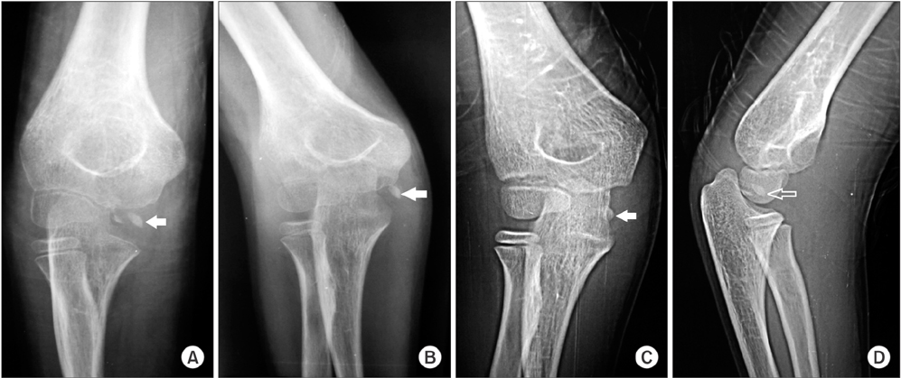
Fig. 1 Preoperative plain radiographs of the right elbow. Note that various degrees of forearm rotation in the anteroposterior (A, B, C) and oblique views (D) affect medial epicondylar fragment visualization (arrows). The depicted epicondylar fragment was shown to be equivalent in size to that of the normal elbow (see Fig. 4E).
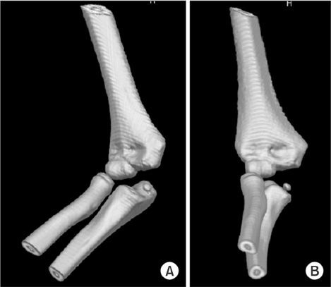
Fig. 2 Three-dimensional volume rendering computed tomography images (A, B) are apparently impressive of intra-articular fragment location. Note widening of the humeroulnar joint space.
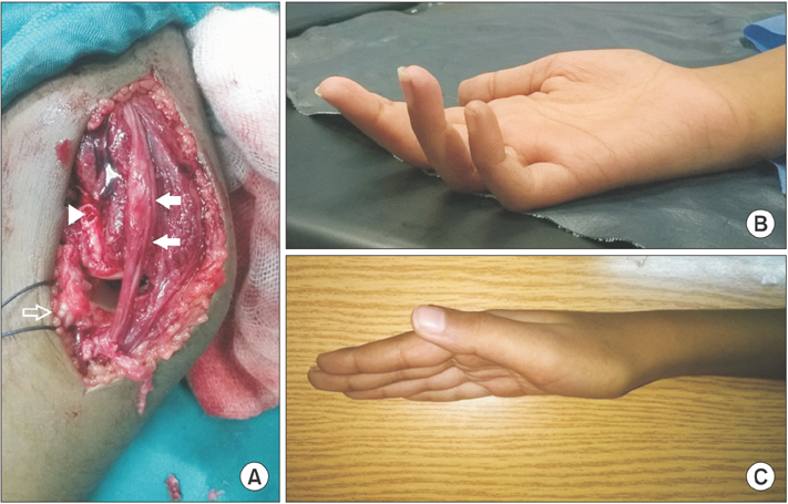
Fig. 3 Ulnar nerve status. (A) Intraoperative image. Note the contused segment (arrows) of the ulnar nerve, the retracted stump of the flexor-pronator mass after fragment excision (hollow arrow), and the medial epicondyle fracture bed (arrow head). (B) Preoperative photograph showing ulnar clawing. (C) One-year postoperative photograph demonstrating full resolution.

Fig. 4 One-year follow-up imaging evaluation. Note the irregular fragmented appearance of the trochlear epiphysis (arrows) in the anteroposterior radiograph (A) and axial (B), sagittal (C), and coronal (D) computed tomography images. (E) Anteroposterior radiograph of the normal left elbow at the same follow-up session. Note the ossified medial epicondylar fragment (arrow) and absent trochlear epiphysis ossification. (F) Longitudinal ultrasound images of the right (RT) and left (LT) common flexor origin showing fanning of the right (injured) one due to reattachment.
Reference
-
1. Pathy R, Dodwell ER. Medial epicondyle fractures in children. Curr Opin Pediatr. 2015; 27(1):58–66.
Article2. Gottschalk HP, Eisner E, Hosalkar HS. Medial epicondyle fractures in the pediatric population. J Am Acad Orthop Surg. 2012; 20(4):223–232.
Article3. Papavasiliou VA. Fracture-separation of the medial epicondylar epiphysis of the elbow joint. Clin Orthop Relat Res. 1982; (171):172–174.
Article4. Kamath AF, Baldwin K, Horneff J, Hosalkar HS. Operative versus non-operative management of pediatric medial epicondyle fractures: a systematic review. J Child Orthop. 2009; 3(5):345–357.
Article5. Lima S, Correia JF, Ribeiro RP, et al. A rare case of elbow dislocation associated with unrecognized fracture of medial epicondyle and delayed ulnar neuropathy in pediatric age. J Shoulder Elbow Surg. 2013; 22(3):e9–e11.
Article6. Haflah NH, Ibrahim S, Sapuan J, Abdullah S. An elbow dislocation in a child with missed medial epicondyle fracture and late ulnar nerve palsy. J Pediatr Orthop B. 2010; 19(5):459–461.
Article7. Cusick MC, Bonnaig NS, Azar FM, Mauck BM, Smith RA, Throckmorton TW. Accuracy and reliability of the Mayo Elbow Performance Score. J Hand Surg Am. 2014; 39(6):1146–1150.
Article8. Keats TE, Anderson MW. Atlas of normal roentgen variants that may simulate disease. 9th ed. Philadelphia, PA: Saunders;2012.
- Full Text Links
-
- Actions
-
Cited
- CITED
-
- Close
- Share
-
- Similar articles
-
- Tardy Ulnar Nerve Palsy due to Recurrent Dislocation of the Ulnar Nerve after Medial Epicondylar Fracture of the Humerus: A Case Report
- Operative treatment of fracture of medial epicondyle of humerus in children
- Ulnar nerve palsy After Percutaneous Pinning in Childrens Supracondylar fracture
- An Ipsilateral Crossed Pinning Technique to Fix Supracondylar Fractures of the Humerus in Children: A Report on the Technique to Escape form Ulnar Nerve Injuries
- Anatomical Evaluation of Ulnar Nerve according to the Elbow Position
