Little Leaguer's Shoulder Can Cause Severe Three-Dimensional Humeral Deformity
- Affiliations
-
- 1Doi Orthopaedic Clinic, Takatsuki, Japan.
- 2Department of Orthopedic Surgery, Osaka Medical College, Takatsuki, Japan. tmihata@osaka-med.ac.jp
- 3First Towakai Hospital, Takatsuki, Japan.
- 4Katsuragi Hospital, Kishiwada, Japan.
- KMID: 2412257
- DOI: http://doi.org/10.4055/cios.2017.9.4.537
Abstract
- We analyzed three-dimensional (3D) humeral deformity (valgus-varus, flexion-extension, and rotational deformation) after little leaguer's shoulder using 3D computed tomography in a 15-year-old male baseball player. Humeral retroversion was increased by 27.1° on the dominant side compared with the nondominant side. Compared with the nondominant shaft, the dominant humeral shaft was deformed in the varus direction (9.4°), resulting in a decreased neck-shaft angle (dominant side, 127.5°; nondominant side, 135.1°), and it was also deformed in the extension direction (21.0°). This case demonstrates that little leaguer's shoulder can cause markedly greater humeral retroversion than has been reported previously and can result in varus and extension deformation of the humerus. These findings suggest that humeral deformity in overhead throwing athletes may not always solely reflect adaptation to throwing.
MeSH Terms
Figure
-
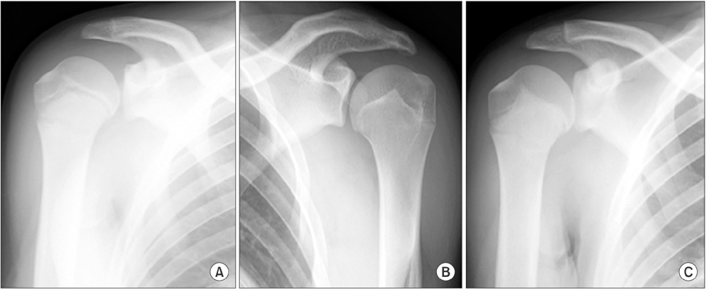
Fig. 1 Plain radiographs in neutral rotation of the patient's shoulder (age, 13 years). (A) Physeal widening in the proximal humerus on the dominant side due to little leaguer's shoulder. (B) Proximal humerus on the nondominant side. (C) After 2 months of rest from throwing, the appearance of the proximal physis became normal and the patient was able to resume throwing without shoulder pain.

Fig. 2 Three-dimensional surface models of the dominant (blue) and nondominant (white) proximal humeri. (A) Anterior view. (B) Lateral view. (C) Posterior view.

Fig. 3 Three-dimensional surface models of the dominant (blue) and nondominant (white) proximal humeri superior to the physis. Anterior (A), lateral (B), posterior (C), superior (D), and inferior (E) views.

Fig. 4 Three-dimensional surface models of the dominant (blue) and nondominant (white) proximal humeri. (A) Posterior view. Humeral retroversion was increased by 27.1° on the dominant side compared with the nondominant side when the image of the dominant humeral shaft was superimposed on the corresponding mirror image of the nondominant side. (B) Anterior view. The dominant humeral shaft was deformed in the varus direction by 9.4° when the image of the dominant humeral head was superimposed on the corresponding mirror image of the nondominant side. (C) Lateral view. The dominant humeral shaft was deformed in the extension direction by 21.0° when the image of the dominant humeral head was superimposed on the corresponding mirror image of the nondominant side.
Reference
-
1. Dotter WE. Little leaguer's shoulder: a fracture of the proximal epiphysial cartilage of the humerus due to baseball pitching. Guthrie Clin Bull. 1953; 23(1):68–72.2. Fleming JL, Hollingsworth CL, Squire DL, Bisset GS. Little Leaguer's shoulder. Skeletal Radiol. 2004; 33(6):352–354.
Article3. Itami Y, Mihata T, Shibano K, Sugamoto K, Neo M. Site and severity of the increased humeral retroversion in symptomatic baseball players: a 3-dimensional computed tomographic analysis. Am J Sports Med. 2016; 44(7):1825–1831.
Article4. Crockett HC, Gross LB, Wilk KE, et al. Osseous adaptation and range of motion at the glenohumeral joint in professional baseball pitchers. Am J Sports Med. 2002; 30(1):20–26.
Article5. Mihata T, Takeda A, Kawakami T, et al. Isolated glenohumeral range of motion, excluding side-to-side difference in humeral retroversion, in asymptomatic high-school baseball players. Knee Surg Sports Traumatol Arthrosc. 2016; 24(6):1911–1917.
Article6. Bishop JY, Flatow EL. Pediatric shoulder trauma. Clin Orthop Relat Res. 2005; (432):41–48.
Article7. Fujisawa Y, Mihata T, Murase T, Sugamoto K, Neo M. Three-dimensional analysis of acromial morphologic characteristics in patients with and without rotator cuff tears using a reconstructed computed tomography model. Am J Sports Med. 2014; 42(11):2621–2626.
Article8. Kohler R, Trillaud JM. Fracture and fracture separation of the proximal humerus in children: report of 136 cases. J Pediatr Orthop. 1983; 3(3):326–332.9. Bahrs C, Zipplies S, Ochs BG, et al. Proximal humeral fractures in children and adolescents. J Pediatr Orthop. 2009; 29(3):238–242.
Article10. Anz AW, Bushnell BD, Griffin LP, Noonan TJ, Torry MR, Hawkins RJ. Correlation of torque and elbow injury in professional baseball pitchers. Am J Sports Med. 2010; 38(7):1368–1374.
Article
- Full Text Links
-
- Actions
-
Cited
- CITED
-
- Close
- Share
-
- Similar articles
-
- Chronic locked anterior shoulder dislocation with impaction of the humeral head onto the coracoid: a case report
- Ultrasonographic Findings of Little Leaguer's Shoulder
- Radiologic Results of Three-Dimensional Templating for Total Shoulder Arthroplasty
- Rapidly Progressive Osteonecrosis of the Humeral Head after Arthroscopic Bankart and Rotator Cuff Repair in a 66-Year Old Woman: A Case Report
- Dislocation of the Shoulder with Ipsilateral Humeral Shaft Fracture: A Case Report
