Two Cases of Cor Triatriatum Associated with Acute Onset Dyspnea in Old Age
- Affiliations
-
- 1Department of Internal Medicine, WonKwang University Medical College, Iksan, Korea.
- KMID: 2410411
- DOI: http://doi.org/10.4250/jkse.1996.4.1.91
Abstract
- Cor triatriatum is a rare congenital heart disease. In the calssic form the left atrium is divided into two chambers by a fibromuscular diaphragm covered by endocardium. The dorsal chamber receives the pulmonary veins, and the ventral chamber gives rise to the left atrial appendage and leads to the mitral valve. Even though the calssic case of cor triatriatum clinically looks like pulmonary venous obstruction, the association, location, and size of the ASD, as well as other cardiac anomalies, may influence symptoms, making the diagnosis difficult. The diagnosis of cor triatriatum is made chiefly from two-dimensional echocardiography and transesophageal echocardiography. The diagnosis can be established by the surgical exploration. In this paper we will report two cases of non-obstruction or triatriatum and review of literature was made.
Keyword
MeSH Terms
Figure
-
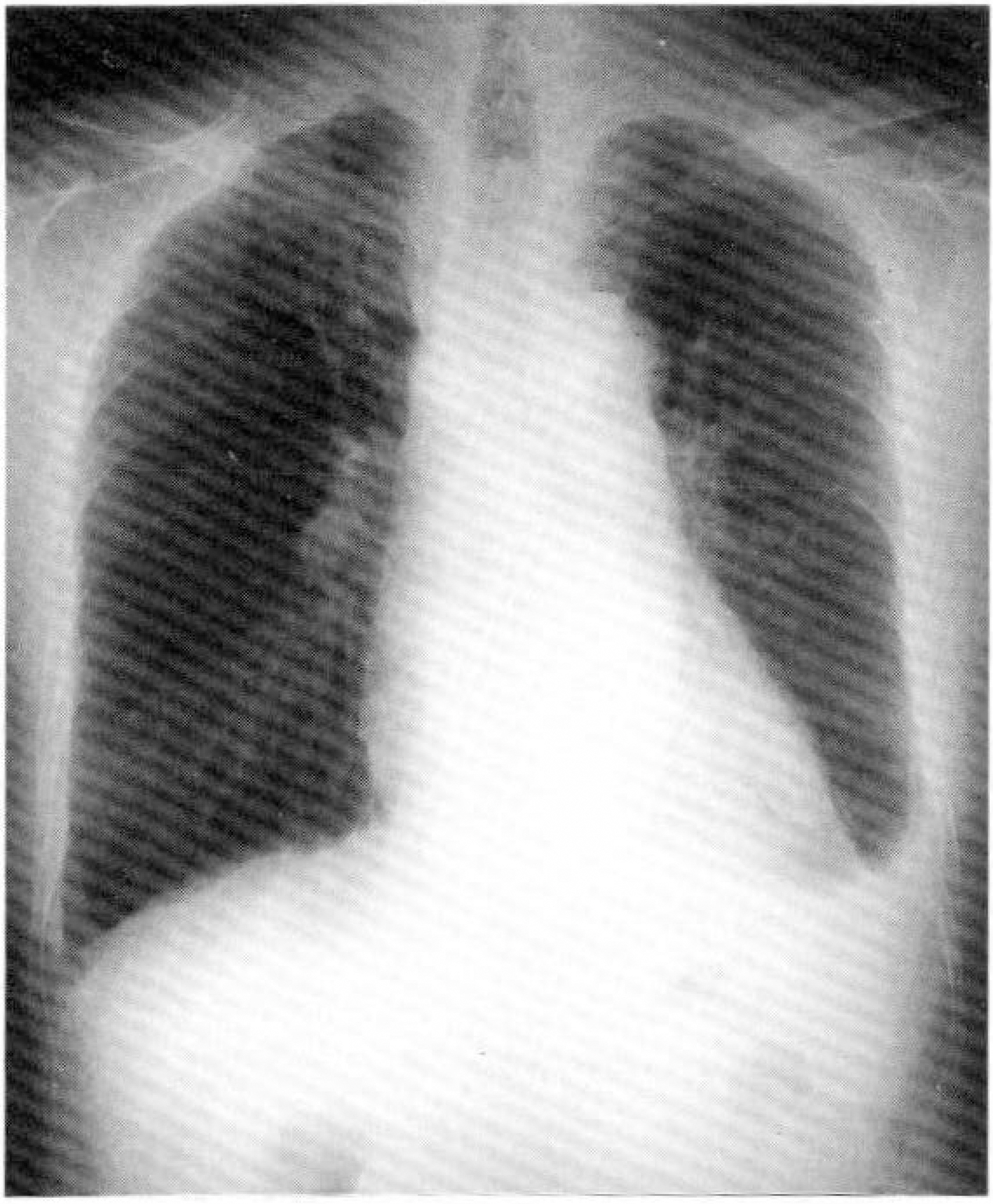
Fig. 1. Chest X-ray shows mild pulmonary vascular congestion and both pleural effusion.
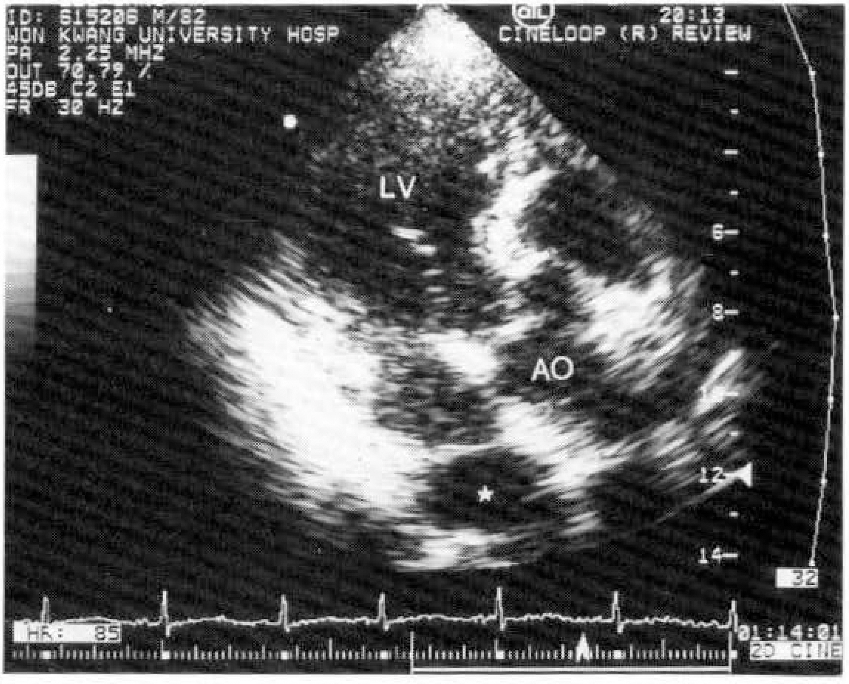
Fig. 2. Five chamber view with the fenestrated intra-atrial membrane in LA represented by transesotphaseal echocardiography. (LV: Left ventricle, AO: Aorta)
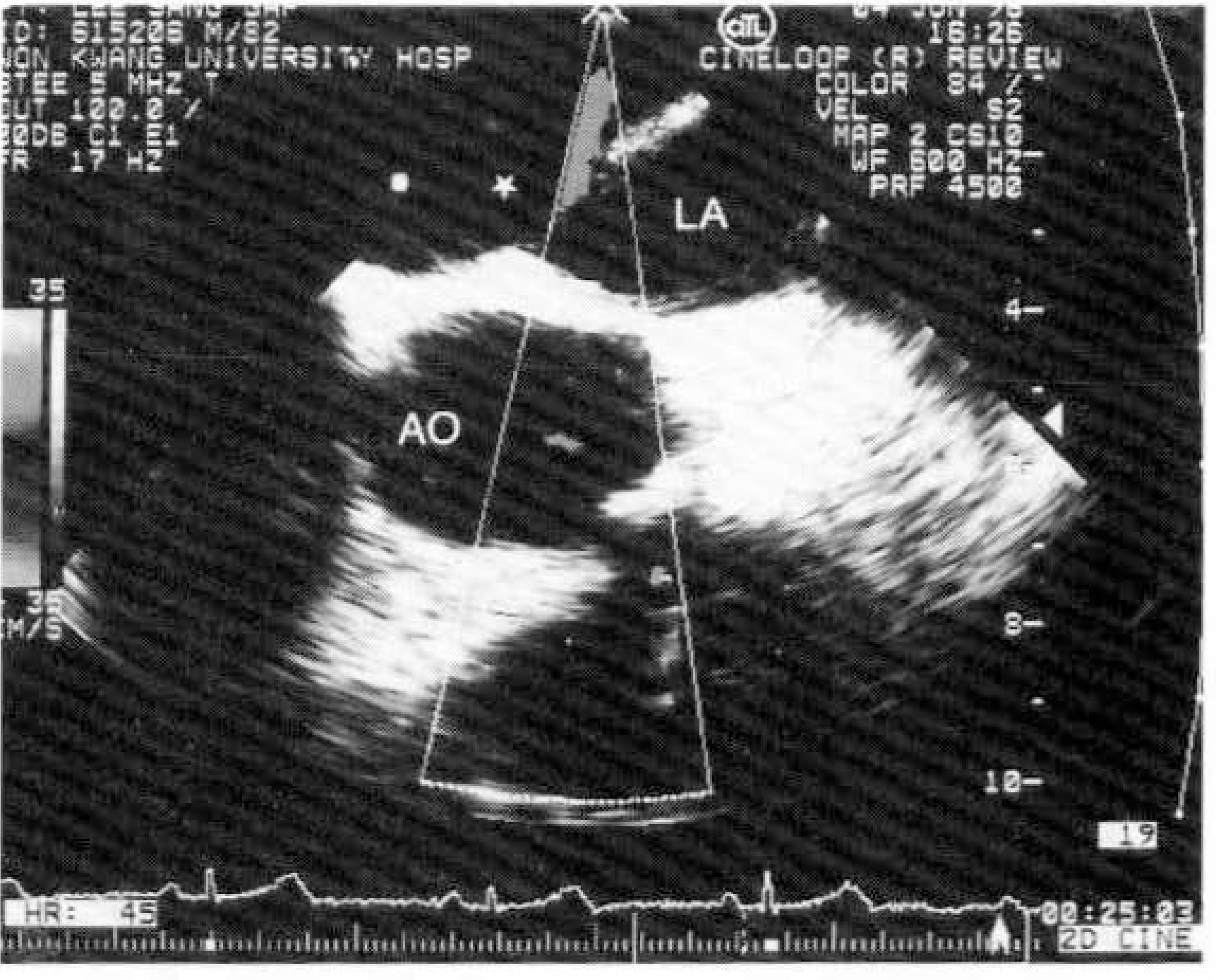
Fig. 3. Basal short axis planes demonstrated by transesophageal echocardiography. Intra-atrial membrane divides the LA into posterosuperior LA and anteroinferior. The opening in the intra-atrial membrane is 2cm sized without obstruction. (LA: Left atrium, AO: Aorta)
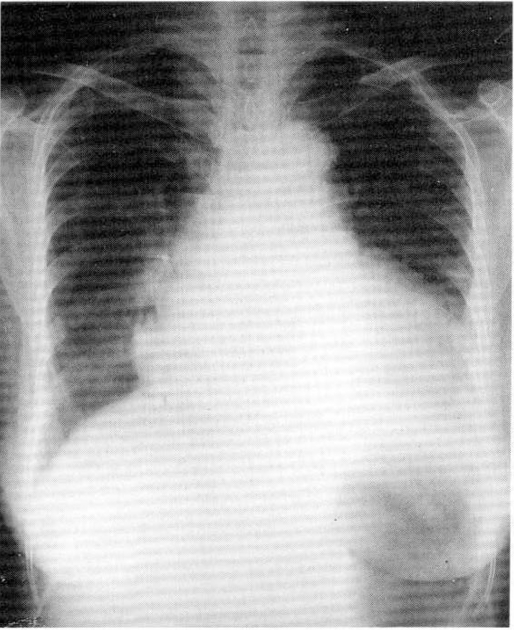
Fig. 4. Chest X-ray shows cardiomegaly and mild pulmonary vascular congestion.
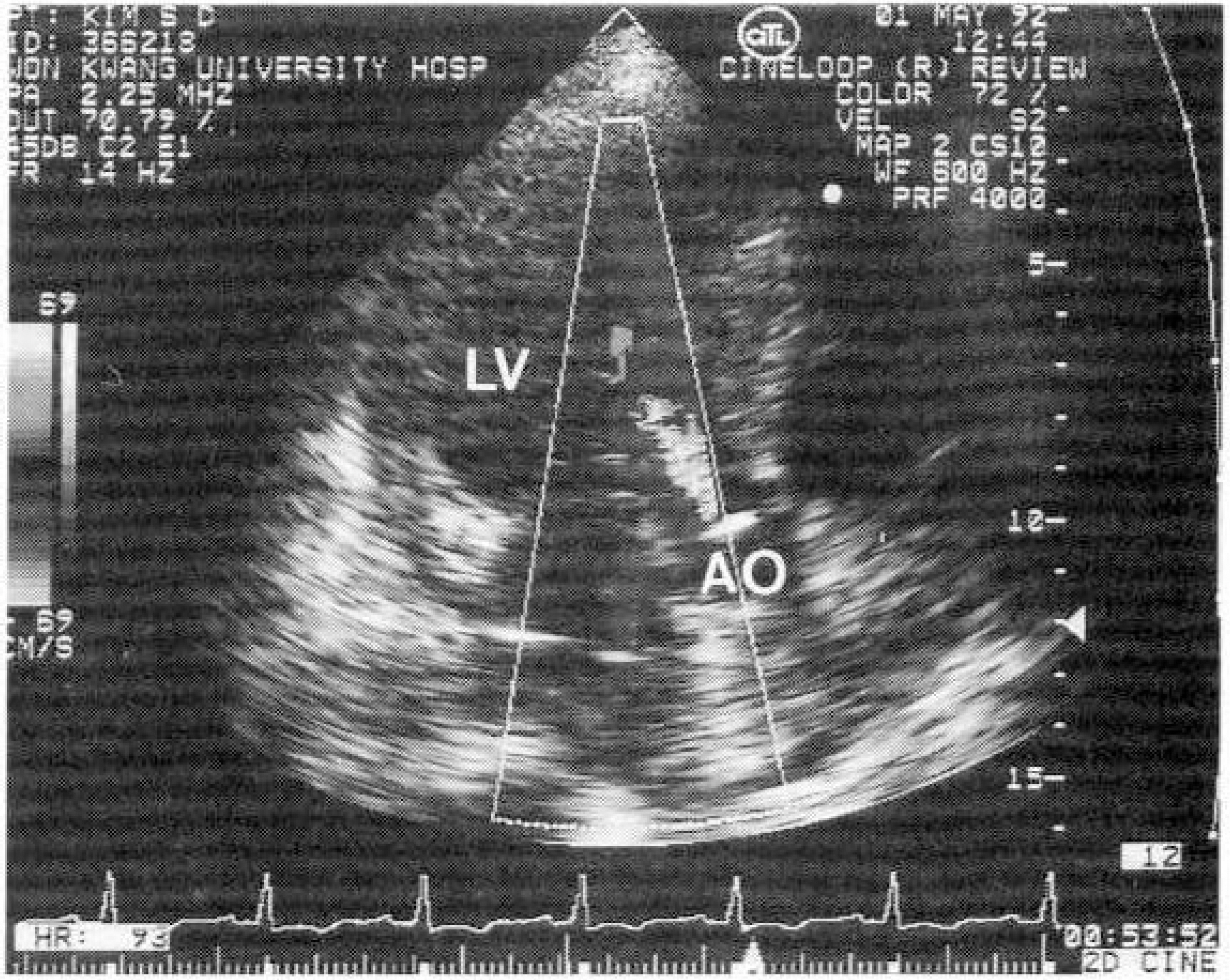
Fig. 5. Five chamber view with color doppler demonstrated transthoracic echocardiography. Intra-atrial membrane in LA was visualized. (LV: Left ventricle, AO: Aorta)
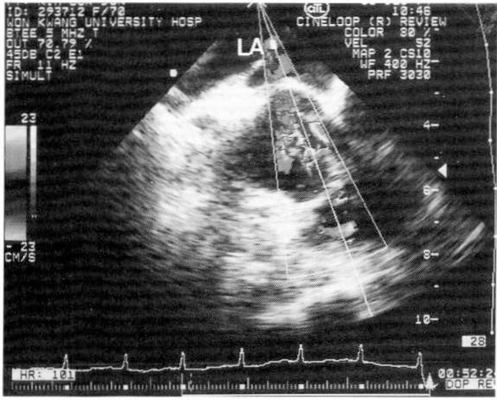
Fig. 6. Basal short axis planes by TEE. Intra-atrial membrane devides the LA into posterosuperior LA and anteroinferior without obstruction. (LA: Left atrium)
Reference
-
References
1). Church WS. Congenital malformation of the heart abnormal septum in left auricle In: Cor triatriatum. Am Heart J. 59:291. 1960.2). 정경영 이두영 · 흉승록, 이-웅구. 삼종방심 치험 l예. 대흉외지. 16:331. 1983.3). 손제분 – 문광덕 이재필 정원상 · 깅영학 – 강정 호 · 지행옥 선정국 ‘ 삼심방증 처l험 2예 대흉외 지. 26:543. 1993.4). 이우승 · 강덕형 – 조성욱 · 김동운 · 손대원 오병 희 이명묵 박영배-최윤식 서정폰·이영우 삼심방싱 l예. 대한내과학회지. 38:257. 1992.5). 김판급 · 이상훈 황홍곤 김영아 – 홍석근 – 후천 성 증상적 승모판협착 환자에게서 발견된 비폐 쇄 성 삼심방심. 경식도 심초음파도 소견‘ 세종의학. JO:217. 1993.6). 김영태 – 노준량. 삼방싱의 외파적 치료; 수술 치 험 24례. 대흉외 지. 27:25끼. 1994.7). Burton DA, Chin A, Weinberg PM, Pigott JP. Identification of cor triatriatum dexter by 2-D echocardiography. Am J Cardiol. 60:409. 1987.8). Alboliras ET, Edward WD, Driscoll DJ, Seward JB. Cor triatriatum dexter: Two-dimentional echocardiographic diagnosis. J Am Coll Cardiol. 9:334. 1987.9). Niwayama G. Cor triatriatum. Am Heart J. 59:291. 1960.10). Jegier W, Gibbons JE, Wigglerworth FW. Cor triatriatum: Clinical hemodynamic and pathologiacal studies. Surgical correction in early life. Pediatrics. 31:255. 1963.11). Griffith TW. Note on a second example of division of the cavity of the left auricle into two compartment by a fibrous band. Cor triatriatum. Am Heart J. 59:291. 1960.12). Van Praagh R, Corsini J. Cor triatriatum: Pathologic anatomy and a consideration of morphogenesis based on 13 postmortem cases and a study of normal development of the pulmonary vein and atrial septum in 83 human embryos. Am Heart J. 78:379. 1969.
Article13). Lucas RV. Congenital causes of pulmonary venous obstruction. Cardiovas Clin. 4:19. 1972.
Article14). Marin-Garcia J, Tandon R, Lucas RV, Edwards JE. Cor triatriatum: Study of 20 cases. Am J Cardiol. 35:59. 1975.15). Patt MV, Obeid AI. Cor triatriatum with isolated pulmonary venous stenosis in an adult: Diagnosis with transesophageal two-dimensional echocardiography. J Am Soc Echo. 4:185. 1991.
Article16). Wong CK, Leung WM, Cheng CH, Lau CP, Cheng DLC. Myxomatous mitral valve degeneration complicating asymptomatic cor triatriatum. Clin Cardiol. 12:48. 1989.
Article17). Gibson DJ, Honey M, Lennox SC. Cor triatriatum diagnosis by echocardiography. British Heart J. 36:835. 1974.
Article18). Canedo MI, Stefadouros MA, Frank MJ, Moore HV, Cundey DW. Echocardiographic feature of cor triatriatum. Am J of Cardiology. 40:615. 1977.19). Oglietti J, Cooley DA, Izquierdo JP, Ventemiglea R, Muasher I, Hallman GL, Reul GJ. Cor triatriatum: Operative results in 25 patients. Ann Thorac Surg. 35:415. 1983.
Article20). Keith JD, Rowe RD, Vlad P. Heart disease in infancy and childhood. 3rd ed.p. 577. 1978.21). Perry LW, Scott LP, McClenathan JE. Cor triatriatum: Preoperative diagnosis and successful surgical repair in a small infant. J of Pediatrics. 71:840. 1967.
