Tarsal Tunnel Syndrome Resulting from a Joint Originated the Intraneural Ganglion of the Medial Plantar Nerve: A Case Report: Surgical Treatment for Prevention of Recurrence
- Affiliations
-
- 1Department of Orthopedic Surgery, Kyung Hee University Hospital at Gangdong, Seoul, Korea.
- 2Department of Orthopedic Surgery, Yeungnam University Hospital, Daegu, Korea. chpark77@naver.com
- KMID: 2407375
- DOI: http://doi.org/10.14193/jkfas.2018.22.1.44
Abstract
- There are a few reports on tarsal tunnel syndrome resulting from the intraneural ganglion. Although it can occur through a connection with the adjacent joint, there is no consensus on its pathogenesis and treatment method. This paper reports a case of tarsal tunnel syndrome resulting from the intraneural ganglion of the medial plantar nerve of the tibial nerve.
Figure
-
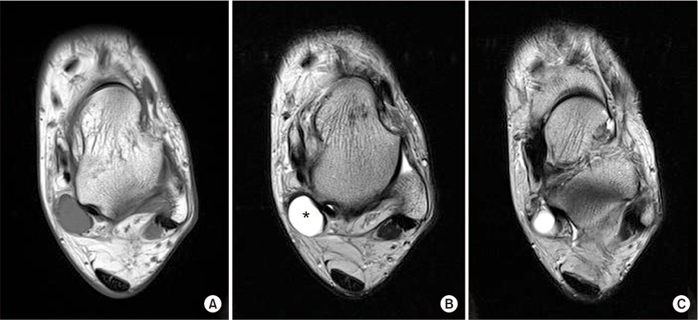
Figure 1 (A, B) Magnetic resonance images showed a 1.5×1.5-cm-sized cystic mass around the medial plantar nerve with homogeneous low signal intensity on the T1-weighted image and homogeneous high signal intensity on T2-weighted image. (C) Axial T2 image was suspected as an articular branch of intraneural ganglion connecting to the adjacent subtalar joint. Asterisk, medial plantar nerve intraneural ganglion.
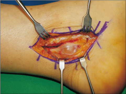
Figure 2 The cystic mass was located in the epineurium of the medial plantar nerve.
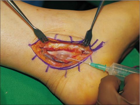
Figure 3 The cystic mass was aspirated using a syringe, following the decompressed nerve was identified.
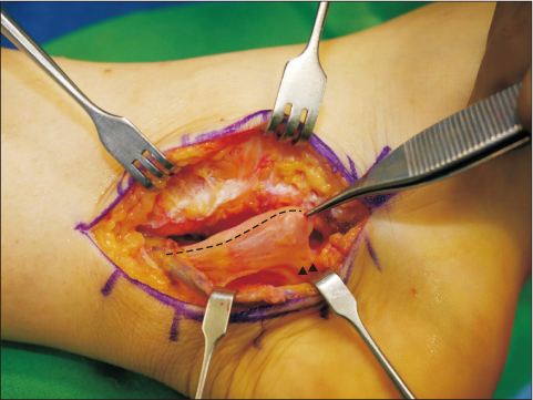
Figure 4 After the aspiration, as shown in the magnetic resonance images, cystic mass and its articular branch connecting to the adjacent subtalar joint were identified. Dashed line, decompressed medial plantar nerve; black arrowheads, articular branch.

Figure 5 Connection between cystic mass and adjacent subtalar joint was identified behind the sustentaculum tali (white arrowheads). Meticulous debridement of the involved joint capsule and flexor halluces tendon sheath was performed.
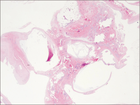
Figure 6 Photomicrograph of the resected lesion was diagnosed as a ganglion with the cyst wall consisted of the degenerative collagen fiber containing mucinous material (H&E stain, ×100).
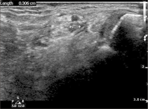
Figure 7 At 18 months after surgery, no recurrence of ganglion was observed in medial plantar nerve on ultrasonography.
Reference
-
1. Ahmad M, Tsang K, Mackenney PJ, Adedapo AO. Tarsal tunnel syndrome: a literature review. Foot Ankle Surg. 2012; 18:149–152.
Article2. Lau JT, Daniels TR. Tarsal tunnel syndrome: a review of the literature. Foot Ankle Int. 1999; 20:201–209.
Article3. Sung KS, Park SJ. Short-term operative outcome of tarsal tunnel syndrome due to benign space-occupying lesions. Foot Ankle Int. 2009; 30:741–745.
Article4. Spinner RJ, Desy NM, Rock MG, Amrami KK. Peroneal intraneural ganglia. Part I. Techniques for successful diagnosis and treatment. Neurosurg Focus. 2007; 22:E16.5. Spinner RJ, Wang H. The first described joint-associated intraneural ganglion cyst. Neurosurgery. 2011; 69:1291–1298.
Article6. Fujita I, Matsumoto K, Minami T, Kizaki T, Akisue T, Yamamoto T. Tarsal tunnel syndrome caused by epineural ganglion of the posterior tibial nerve: report of 2 cases and review of the literature. J Foot Ankle Surg. 2004; 43:185–190.
Article7. Spinner RJ, Dellon AL, Rosson GD, Anderson SR, Amrami KK. Tibial intraneural ganglia in the tarsal tunnel: is there a joint connection? J Foot Ankle Surg. 2007; 46:27–31.
Article8. Friedlander HL. Intraneural ganglion of the tibial nerve. A case report. J Bone Joint Surg Am. 1967; 49:519–522.9. Spinner RJ, Atkinson JL, Harper CM Jr, Wenger DE. Recurrent intraneural ganglion cyst of the tibial nerve. Case report. J Neurosurg. 2000; 92:334–337.10. Chick G, Alnot JY, Silbermann-Hoffman O. Benign solitary tumors of the peripheral nerves. Rev Chir Orthop Reparatrice Appar Mot. 2000; 86:825–834.
- Full Text Links
-
- Actions
-
Cited
- CITED
-
- Close
- Share
-
- Similar articles
-
- Ganglion of Flexor Digitorum Longus Tendon Sheath and Multifocal Myxoid Degeneration of Medial Plantar Nerve Producing Tarsal Tunnel Syndrome: A Case Report
- Surgical Decompression of Tarsal Tunnel Syndrome Associated with Ganglion Cyst: A Case Report
- Cubital tunnel syndrome caused by an intraneural ganglion cyst treated with epineurectomy: a report of three cases
- Tarsal Tunnel Syndrome caused by Neurilemoma of the Posterior Tibial Nerve: A case Report
- Intraneural Ganglion of Ulnar Nerve in Proximal Forearm: Case Report
