Estimation of the effective dose of dental cone-beam computed tomography using personal computer-based Monte Carlo software
- Affiliations
-
- 1Department of Oral and Maxillofacial Radiology, College of Dentistry, Dankook University, Cheonan, Korea. ekkim@dankook.ac.kr
- 2Dental Hospital, School of Dentistry, Mongolian National University of Medical Sciences, Ulaanbaatar, Mongolia.
- KMID: 2406979
- DOI: http://doi.org/10.5624/isd.2018.48.1.21
Abstract
- PURPOSE
To calculate the effective doses of cone-beam computed tomography (CBCT) using personal computer-based Monte Carlo (PCXMC) software (Radiation and Nuclear Safety Authority, Helsinki, Finland) and to compare the calculated effective doses with those measured using thermoluminescent dosimeters (TLDs) and an anthropomorphic phantom.
MATERIALS AND METHODS
An Alphard VEGA CBCT scanner (Asahi Roentgen Ind. Co., Kyoto, Japan) with multiple fields of view (FOVs) was used for this study. The effective doses of the scout and main projections of CBCT using 1 large and 2 medium FOVs with a height >10 cm were calculated using PCXMC and PCXMCRotation software and then were compared with the doses obtained using TLD-100 LiF and an anthropomorphic adult human male phantom. Furthermore, it was described how to determine the reference points on the Y- and Z-axes in PCXMC, the important dose-determining factors in this software.
RESULTS
The effective doses at CBCT for 1 large (20.0 cm×17.9 cm) and 2 medium FOVs (15.4 cm×15.4 cm and 10.2 cm×10.2 cm) calculated by the PCXMC software were 181, 300, and 158 µSv, respectively. These values were comparable (16%-18% smaller) to those obtained through TLD measurements in each mode.
CONCLUSION
The use of PCXMC software could be an alternative to the TLD measurement method for effective dose estimation in CBCT with large and medium FOVs.
Figure
-

Fig. 1 Coronal image of 3-dimensionally reconstructed cone-beam computed tomography (C mode) of the phantom with superimposed PCXMC Z coordinates. The Zref of the rotation center was set to approximately 86.2 cm.

Fig. 2 Coronal image of 3-dimensionally reconstructed cone-beam computed tomography (P mode) of the phantom with superimposed PCXMC Z coordinates. The Zref of the rotation center was set to approximately 85.0 cm.

Fig. 3 Coronal image of 3-dimensionally reconstructed cone-beam computed tomography (I mode) of the phantom with superimposed PCXMC Z coordinates. The Zref of the rotation center was set to approximately 84.5 cm.
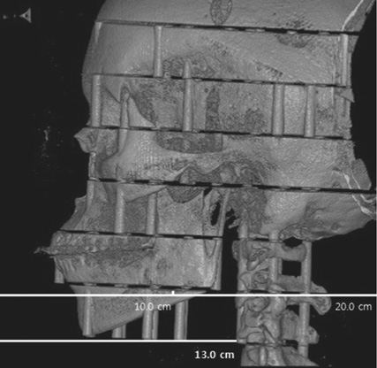
Fig. 4 Lateral image of 3-dimensionally reconstructed cone-beam computed tomography (C mode) of the phantom. The width of this image is 20.0 cm and the length from the left margin to most mid-anterior point of the cervical vertebrae is 13.0 cm. Thus, the length from center to most mid-anterior point of cervical vertebrae is 3.0 cm. Meanwhile, the Y-axis value of the most mid-anterior point of cervical vertebrae inferior to the mandible was approximately −1.0. Thus, the Yref value of the rotation center was determined to be approximately −4.0 cm.

Fig. 5 Lateral image of 3-dimensionally reconstructed cone-beam computed tomography (P mode) of the phantom. The width of this image is 15.4 cm and the length from the left margin to most mid-anterior point of the cervical vertebrae is 10.5 cm. Thus, the length from center to most mid-anterior point of cervical vertebrae is 2.8 cm and the Yref value of the rotation center was determined to be approximately −3.8 cm.

Fig. 6 Lateral image of 3-dimensionally reconstructed cone-beam computed tomography (I mode) of the phantom. The width of this image is 10.2 cm and the length from the left margin to most mid-anterior point of the cervical vertebrae is 9.9 cm. Thus, the length from center to most mid-anterior point of cervical vertebrae is 4.8 cm and the Yref value of the rotation center was determined to be approximately −5.8 cm.
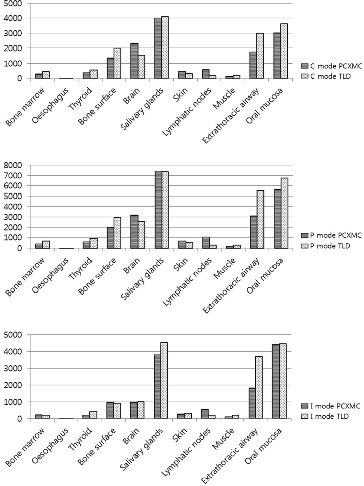
Fig. 7 Bar graphs show the absorbed organ doses in various organs and tissues calculated by PCXMC software and measured using TLD technology (Upper: C mode, Middle: P mode for maxilla, Lower: I mode for maxilla). C: cephalometric, P: panoramic, I: implant, TLD: thermoluminescent dosimetry.
Reference
-
1. Kim DS, Rashsuren O, Kim EK. Conversion coefficients for the estimation of effective dose in cone-beam CT. Imaging Sci Dent. 2014; 44:21–29.
Article2. Ludlow JB, Timothy R, Walker C, Hunter R, Benavides E, Samuelson D, et al. Effective dose of dental CBCT - a meta analysis of published data and additional data for nine CBCT units. Dentomaxillofac Radiol. 2015; 44:20140197.3. Martin CJ. Effective dose: how should it be applied to medical exposures? Br J Radiol. 2007; 80:639–647.
Article4. Pauwels R, Beinsberger J, Collaert B, Theodorakou C, Rogers J, Walker A, et al. Effective dose range for dental cone beam computed tomography scanners. Eur J Radiol. 2012; 81:267–271.
Article5. Al-Okshi A, Lindh C, Salé H, Gunnarsson M, Rohlin M. Effective dose of cone beam CT (CBCT) of the facial skeleton: a systematic review. Br J Radiol. 2015; 88:20140658.
Article6. Ludlow JB, Ivanovic M. Comparative dosimetry of dental CBCT devices and 64-slice CT for oral and maxillofacial radiology. Oral Surg Oral Med Oral Pathol Oral Radiol Endod. 2008; 106:106–114.
Article7. Koivisto J, Kiljunen T, Tapiovaara M, Wolff J, Kortesniemi M. Assessment of radiation exposure in dental cone-beam computerized tomography with the use of metal-oxide semiconductor field-effect transistor (MOSFET) dosimeters and Monte Carlo simulations. Oral Surg Oral Med Oral Pathol Oral Radiol. 2012; 114:393–400.
Article8. Aps JK, Scott JM. Oblique lateral radiographs and bitewings; estimation of organ doses in head and neck region with Monte Carlo calculations. Dentomaxillofac Radiol. 2014; 43:20130419.
Article9. Lee C, Lee SS, Kim JE, Huh KH, Yi WJ, Heo MS, et al. Comparison of dosimetry methods for panoramic radiography: thermoluminescent dosimeter measurement versus personal computer-based Monte Carlo method calculation. Oral Surg Oral Med Oral Pathol Oral Radiol. 2016; 121:322–329.
Article10. Podnieks EC, Negus IS. Practical patient dosimetry for partial rotation cone beam CT. Br J Radiol. 2012; 85:161–167.
Article11. Vassileva J, Stoyanov D. Quality control and patient dosimetry in dental cone beam CT. Radiat Prot Dosimetry. 2010; 139:310–312.
Article12. Bai M, Liu X, Liu B. Effective patient dose during neuroradiological C-arm CT procedures. Diagn Interv Radiol. 2013; 19:29–32.13. He W, Huda W, Magill D, Tavrides E, Yao H. Patient doses and projection angle in cone beam CT. Med Phys. 2010; 37:2359–2368.
Article14. Khelassi-Toutaoui N, Berkani Y, Tsapaki V, Toutaoui AE, Merad A, Frahi-Amroun A, et al. Experimental evaluation of PCXMC and prepare codes used in conventional radiology. Radiat Prot Dosimetry. 2008; 131:374–378.
Article15. Kawasaki T, Aoyama T, Yamauchi-Kawaura C, Fujii K, Koyama S. Organ dose and effective dose estimation in paediatric chest radiographic examinations by using pin silicon photodiode dosemeters. Radiat Prot Dosimetry. 2013; 154:314–319.
Article16. Okano T, Matsuo A, Gotoh K, Yokoi M, Hirukawa A, Okumura S, et al. Comparison of absorbed and effective dose from two dental cone beam computed tomography scanners. Nihon Hoshasen Gijutsu Gakkai Zasshi. 2012; 68:216–225.
Article17. Reeves TE, Mah P, McDavid WD. Deriving Hounsfield units using grey levels in cone beam CT: a clinical application. Dentomaxillofac Radiol. 2012; 41:500–508.
Article18. The 2007 Recommendations of the International Commission on Radiological Protection. ICRP publication 103. Ann ICRP. 2007; 37:1–332.19. Tapiovaara M, Siiskonen T. PCXMC, a Monte Carlo program for calculating patient doses in medical X-ray examinations. 2nd ed. Helsinki: Finnish Centre for Radiation and Nuclear Safety Authority;2008. Report STUK-A231.20. Qu XM, Li G, Ludlow JB, Zhang ZY, Ma XC. Effective radiation dose of ProMax 3D cone-beam computerized tomography scanner with different dental protocols. Oral Surg Oral Med Oral Pathol Oral Radiol Endod. 2010; 110:770–776.
Article
- Full Text Links
-
- Actions
-
Cited
- CITED
-
- Close
- Share
-
- Similar articles
-
- Analysis of Beam Hardening of Modulation Layers for Dual Energy Cone-beam CT
- A Monte Carlo Simulation Study of a Therapeutic Proton Beam Delivery System Using the Geant4 Code
- Management of root canal perforation by using cone-beam computed tomography
- Verification of the PMCEPT Monte Carlo dose Calculation Code for Simulations in Medical Physics
- New evolution of cone-beam computed tomography in dentistry: Combining digital technologies
