Solitary Bladder Metastasis of Prostate Cancer Mimicking Bladder Submucosal Tumor: A Case Report
- Affiliations
-
- 1Department of Radiology, Jeju National University Hospital, Jeju National University School of Medicine, Jeju, Korea. shinshlee@naver.com
- 2Department of Urology, Jeju National University Hospital, Jeju National University School of Medicine, Jeju, Korea.
- 3Department of Pathology, Jeju National University Hospital, Jeju National University School of Medicine, Jeju, Korea.
- KMID: 2405736
- DOI: http://doi.org/10.3348/jksr.2017.77.3.197
Abstract
- Prostate cancer is one of the most common causes of secondary cancer to the bladder. There have been few reports about distant metastasis to the bladder from primary prostate cancer, since secondary involvement of the urinary bladder in prostate cancer is most often by direct invasion. Metastatic prostate cancer to the bladder is often mistaken for other primary bladder tumors. Here, we report a case of solitary metastatic prostatic cancer to the bladder, which was previously misdiagnosed as a submucosal tumor of the bladder.
MeSH Terms
Figure
-
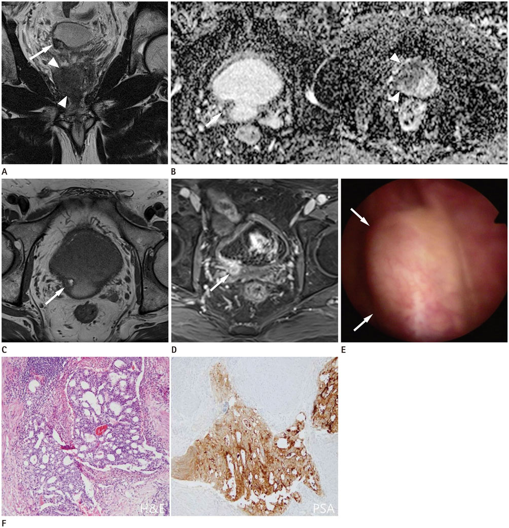
Fig. 1 A Solitary bladder metastasis of prostate cancer in a 75-year-old man. A. The coronal T2-weighted MR image shows a homogeneously moderate hypointense mass in the right lobe of the prostate gland (arrowheads), and a 1.1 cm sized submucosal tumor with heterogeneously high signal intensity at the right posterior wall of the urinary bladder (arrow). There is no evidence of connection between the two lesions. B. Axial apparent diffusion coefficient maps show a heterogeneously moderate hypointense tumor in the urinary bladder (arrow), and a markedly hypointense tumor in the right lobe of the prostate gland (arrowheads). C. Axial T1-weighted MR image shows an isointense tumor with an internal hyperintense portion in the urinary bladder (arrow). D. Contrasted enhanced fat-saturated T1-weighted MR image shows a heterogeneous enhancing tumor in the urinary bladder (arrow). E. Cystoscopy reveals a small submucosal tumor in the right posterior wall of the urinary bladder (arrows). F. Transurethral resection of the bladder tumor histopathologically confirms it as a metastatic prostate adenocarcinoma, which forms multiple cribriform irregular nests infiltrating the muscle layer of the urinary bladder (hematoxylin-eosin stain, original magnification × 100). The tumor cells are strongly positive to prostate-specific antigen (immunohistochemical stain, original magnification × 100).
Reference
-
1. Bates AW, Baithun SI. Secondary neoplasms of the bladder are histological mimics of nontransitional cell primary tumours: clinicopathological and histological features of 282 cases. Histopathology. 2000; 36:32–40.2. Melicow MM. Tumors of the urinary bladder: a clinico-pathological analysis of over 2500 specimens and biopsies. J Urol. 1955; 74:498–521.3. Bubendorf L, Schöpfer A, Wagner U, Sauter G, Moch H, Willi N, et al. Metastatic patterns of prostate cancer: an autopsy study of 1,589 patients. Hum Pathol. 2000; 31:578–583.4. Ganem EJ, Batal JT. Secondary malignant tumors of the urinary bladder metastatic from primary foci in distant organs. J Urol. 1956; 75:965–972.5. Hallemeier CL, Kohli M, Chandan VS, Miller RC, Choo R. Multiple urinary bladder masses from metastatic prostate adenocarcinoma. Rare Tumors. 2010; 2:e65.6. Buchholz NP, Moch H, Mihatsch MJ. Late urinary bladder metastases after radical resection of prostatic carcinoma. Urol Int. 1994; 53:50–52.7. Shirakawa H, Kozakai N, Sugiura H, Hara S. [Urinary bladder metastasis of prostate cancer: a case report]. Hinyokika Kiyo. 2010; 56:123–125.8. Chung AD, Schieda N, Flood TA, Cagiannos I, Kielar AZ, McInnes MD, et al. Suburothelial and extrinsic lesions of the urinary bladder: radiologic and pathologic features with emphasis on MR imaging. Abdom Imaging. 2015; 40:2573–2588.9. Wong-You-Cheong JJ, Woodward PJ, Manning MA, Sesterhenn IA. From the archives of the AFIP: neoplasms of the urinary bladder: radiologic-pathologic correlation. Radiographics. 2006; 26:553–580.10. Torenbeek R, Lagendijk JH, Van Diest PJ, Bril H, van de Molengraft FJ, Meijer CJ. Value of a panel of antibodies to identify the primary origin of adenocarcinomas presenting as bladder carcinoma. Histopathology. 1998; 32:20–27.
- Full Text Links
-
- Actions
-
Cited
- CITED
-
- Close
- Share
-
- Similar articles
-
- Primary Adenocarcinoma of the Bladder Treated by Pelvic Exenteration : Report of A Case
- A case of metastatic adenocarcinoma of bladder from stomach cancer
- Ectopic Prostate Tissue in the Posterior Wall of the Bladder
- The significance of random mucosal biopsy in bladder cancer
- Sudden hypothermia after bladder perforation during transurethral bladder tumor resection: A case report
