Korean J Crit Care Med.
2017 Nov;32(4):370-371. 10.4266/kjccm.2017.00290.
A Pleural Catheter Malposition through Diaphragm to Abdominal Cavity
- Affiliations
-
- 1Division of Respiratory and Critical Care Medicine, Department of Internal Medicine, Korea University Anam Hospital, Korea University College of Medicine, Seoul, Korea. wonjai21@gmail.com
- KMID: 2405125
- DOI: http://doi.org/10.4266/kjccm.2017.00290
Abstract
- No abstract available.
MeSH Terms
Figure
-
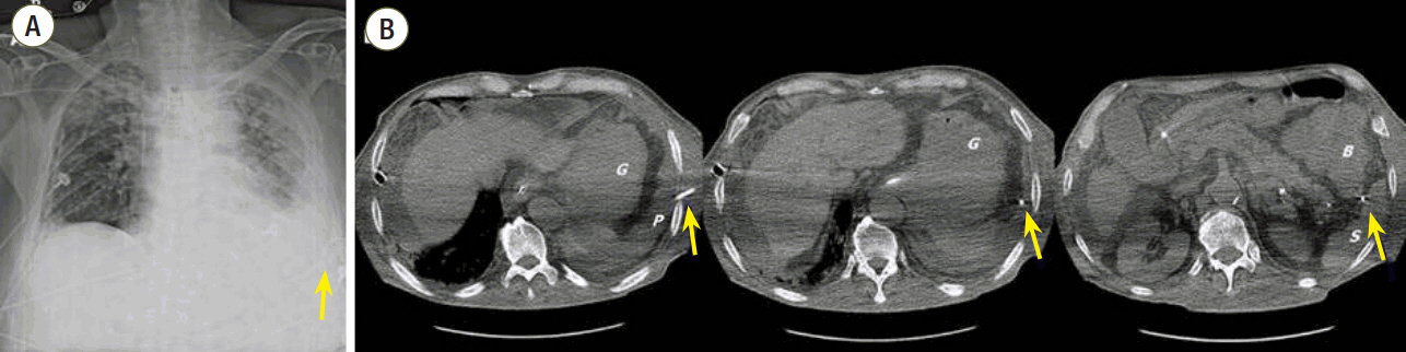
Figure 1. (A) Chest radiography showed the pleural catheter directed downward, marked with arrow. (B) Computed tomography scan showed that the catheter first entered into the pleural space, passed through diaphragm and the tip was located in the abdominal cavity, marked with arrows. G: stomach; P: pleural effusion; B: bowel; S: spleen.
Reference
-
References
1. Havelock T, Teoh R, Laws D, Gleeson F; BTS Pleural Disease Guideline Group. Pleural procedures and thoracic ultrasound: British Thoracic Society Pleural Disease Guideline 2010. Thorax. 2010; 65 Suppl 2:ii61–76.
Article2. Remérand F, Luce V, Badachi Y, Lu Q, Bouhemad B, Rouby JJ. Incidence of chest tube malposition in the critically ill: a prospective computed tomography study. Anesthesiology. 2007; 106:1112–9.3. Kirschbaum A, Damanakis A, Rolfes C, Bartsch D. Endobronchial malposition of matthys pleural catheter: a case report. Thorac Cardiovasc Surg Rep. 2015; 4:14–7.4. Kwiatt M, Tarbox A, Seamon MJ, Swaroop M, Cipolla J, Allen C, et al. Thoracostomy tubes: a comprehensive review of complications and related topics. Int J Crit Illn Inj Sci. 2014; 4:143–55.
Article
- Full Text Links
-
- Actions
-
Cited
- CITED
-
- Close
- Share
-
- Similar articles
-
- Hemothorax induced by postoperative abdominal bleeding in gynecologic patient with undiagnosed porous diaphragm syndrome: A case report
- A Case of Meigs' Syndrome
- A Case of Acute Pancreatitis with Right-sided Pleural Effusion Probably Formed by Aberrant Lymphatic Transfer to the Right Hemidiaphragm
- The Laparoscopic Repair of a Morgagni Hernia in a Child
- Hemothorax Without Injury of the Pleural Cavity due to Diaphragmatic and Liver Laceration Caused by a Right Upper Anterior Chest Stab Wound
