Intraparenchymal Atypical Meningioma in Basal Ganglia Region in a Child: Case Report and Literature Review
- Affiliations
-
- 1Department of Neurosurgery, West China Hospital, Sichuan University, Chengdu City, Sichuan Province, China. 1229013105@qq.com
- KMID: 2403532
- DOI: http://doi.org/10.3340/jkns.2015.0609.001
Abstract
- Intraparenchymal meningiomas without dural attachment are extremely rare, especially when they occur in basal ganglia region in child. An 8-year-old boy was admitted at our hospital, complaining of recurrent headache and vomiting for 3 months. Neurological examination showed impaired vision and mild paresis of the left extremities. Magnetic resonance imaging demonstrated a lesion located in the right basal ganglia region extending to superasellar cistern with solid, multiple cystic and necrotic components. Computed tomography revealed calcification within the mass. Due to the anterior cerebral artery involvement, a subtotal resection was achieved and postoperative radiotherapy was recommended. Histopathological examination indicated that the lesion was an atypical meningioma. The postoperative rehabilitation was uneventful. Mildly impaired vision and motor weakness of left extremities improved significantly and the patient returned to normal life after surgery. To our knowledge, intraparenchymal atypical meningioma in basal ganglia extending to superasellar cistern was never reported. The significance in differential diagnosis of lesions in basal ganglia should be emphasized.
MeSH Terms
Figure
-

Fig. 1 CT revealing calcification, necrosis, cystic formation of the mass lesion (A and B). No hyperostosis or bone absorbtion was observed (C).
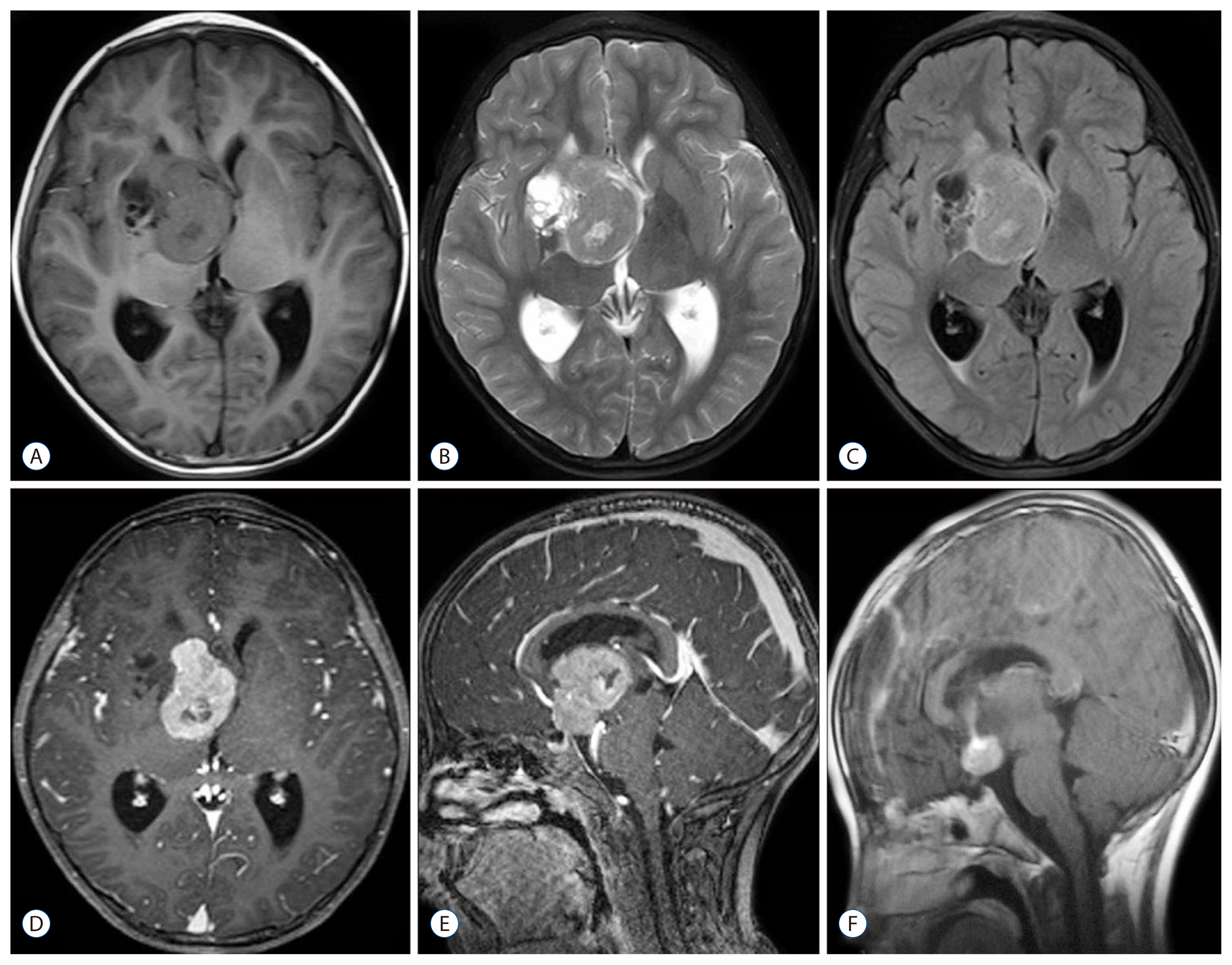
Fig. 2 MRI showing a mass lesion in the right basal ganglia consisted of solid and cystic parts. The solid part manifesting isointense signal on T1-weighted (A), T2-weighted (B), and FLAIR (C) with heterogeneous enhancement (D and E); the cystic part showing hypointense signal on both T1-weighted (A) and FLAIR (C) and hyperintensity on T2-weighted images (B) without enhancement of the wall (D and E). Although the lower margin of the tumor touched the dorsum sellae dura, ‘dura tail’ sign was not noted (E). Postoperative MRI revealing that most of the tumor has been resected and the residual part detached from the dorsum sellae (F). Subdural fluid accumulation in the surgical region was also detected (F). FLAIR: fluid at tenuation inversion recovery, MRI: magetic resonance imaging.

Fig. 3 Hematoxylin-eosin staining exhibiting sheetlike growth, foci of spontaneous, increased cellularity, small cells with a high nucleus-to-cytoplasm ratio and prominent nucleoli (A: original magnification, ×100; B: original magnification, ×400). Immunohistochemical staining showing positive for epitheliod membrane antigen (D: original magnification, ×400), but negative for glial fibrillary acidic protein (E: original magnification, ×400) and S-100 protein (F: original magnification, ×400). Ki-67 labeling index being approximately 10% (C: original magnification, ×400).
Reference
-
References
1. Amirjamshidi A, Mehrazin M, Abbassioun K. Meningiomas of the central nervous system occurring below the age of 17: report of 24 cases not associated with neurofibromatosis and review of literature. Childs Nerv Syst. 16:406–416. 2000.
Article2. Drake JM, Hendrick EB, Becker LE, Chuang SH, Hoffman HJ, Humphreys RP. Intracranial meningiomas in children. Pediatr Neurosci. 12:134–139. 1986.
Article3. Iannelli A, Pieracci N, Bianchi MC, Becherini F, Castagna M. Primary intradiploic meningioma in a child. Childs Nerv Syst. 24:7–11. 2008.
Article4. Im SH, Wang KC, Kim SK, Oh CW, Kim DG, Hong SK, et al. Childhood meningioma: unusual location, atypical radiological findings, and favorable treatment outcome. Childs Nerv Syst. 17:656–662. 2001.
Article5. Higano S, Takahashi S, Ishii K, Matsumoto K, Ikeda H, Sakamoto K. Germinoma originating in the basal ganglia and thalamus: MR and CT evaluation. AJNR Am J Neuroradiol. 15:1435–1441. 1994.6. Jadik S, Stan AC, Dietrich U, Pietilä TA, Elsharkawy AE. Intraparenchymal meningioma mimicking cavernous malformation: a case report and review of the literature. J Med Case Rep. 8:467. 2014.
Article7. Jiang XB, Ke C, Han ZA, Lin SH, Mou YG, Luo RZ, et al. Intraparenchymal papillary meningioma of brainstem: case report and literature review. World J Surg Oncol. 10:10. 2012.
Article8. Jung YS, Song YJ. Meningioma in a 20-month-old boy. J Korean Neurosurg Soc. 51:219–221. 2012.
Article9. Kamoshima Y, Terasaka S, Kobayashi H, Kaneko S, Kubota K, Tanaka S, et al. Radiation induced intraparenchymal meningioma occurring 6 years after CNS germinoma: case report. Clin Neurol Neurosurg. 114:1077–1080. 2012.
Article10. Karadereler S, Aker F, Berkman Z. Intraparenchymal meningioma in a child. Case report and review of the literature. J Neurosurg. 101(1 Suppl):112–115. 2004.11. Kimura H, Nakagawa K, Sakaki S, Matsuoka K. Intracranial meningioma of an infant: a case report. No Shinkei Geka. 15:663–668. 1987.12. Kobayashi T, Yoshida J, Kida Y. Bilateral germ cell tumors involving the basal ganglia and thalamus. Neurosurgery. 24:579–583. 1989.
Article13. Kohama I, Sohma T, Nunomura K, Igarashi K, Ishikawa A. Intraparenchymal meningioma in an infant--case report. Neurol Med Chir (Tokyo). 36:598–601. 1996.14. Kotecha RS, Jacoby P, Cole CH, Gottardo NG. Morbidity in survivors of child and adolescent meningioma. Cancer. 119:4350–4357. 2013.
Article15. Kotecha RS, Junckerstorff RC, Lee S, Cole CH, Gottardo NG. Pediatric meningioma: current approaches and future direction. J Neurooncol. 104:1–10. 2011.
Article16. Kotecha RS, Pascoe EM, Rushing EJ, Rorke-Adams LB, Zwerdling T, Gao X, et al. Meningiomas in children and adolescents: a meta-analysis of individual patient data. Lancet Oncol. 12:1229–1239. 2011.
Article17. Legius E, Vles JS, Casaer P, Plets C, Dom R. Intraparenchymal meningioma in a 14-month-old infant: case report. Brain Dev. 7:622–624. 1985.
Article18. Mamourian AC, Lewandowski AE, Towfighi J. Cystic intraparenchymal meningioma in a child: case report. AJNR Am J Neuroradiol. 12:366–367. 1991.19. Morimoto M, Aoki H, Sadamitsu N, Nakashima R. Cystic meningioma--report of two cases (author’s transl). No Shinkei Geka. 4:805–809. 1976.20. Nayil K, Makhdoomi R, Malik R, Ramzan A. Intraparenchymal anaplastic meningioma in a child: a rare entity. Asian J Neurosurg. 10:111–113. 2015.
Article21. Park I, Huh J, Kim JH, Lee SW, Ryu MH, Kang YK. Primary central nervous system marginal zone B-cell lymphoma of the Basal Ganglia mimicking low-grade glioma: a case report and review of the literature. Clin Lymphoma Myeloma. 8:305–308. 2008.
Article22. Perilongo G, Sutton LN, Goldwein JW, Gusnard D, Schut L, Biegel JA, et al. Childhood meningiomas. Experience in the modern imaging era. Pediatr Neurosurg. 18:16–23. 1992.23. Pinto PS, Huisman TA, Ahn E, Jordan LC, Burger P, Cohen KJ, et al. Magnetic resonance imaging features of meningiomas in children and young adults: a retrospective analysis. J Neuroradiol. 39:218–226. 2012.
Article24. Rasalkar DD, Chu WC, Cheng FW, Paunipagar BK, Shing MK, Li CK. Atypical location of germinoma in basal ganglia in adolescents: radiological features and treatment outcomes. Br J Radiol. 83:261–267. 2010.
Article25. Ravindranath K, Vasudevan MC, Pande A, Symss N. Management of pediatric intracranial meningiomas: an analysis of 31 cases and review of literature. Childs Nerv Syst. 29:573–582. 2013.
Article26. Sakaki S, Nakagawa K, Kimura H, Ohue S. Intracranial meningiomas in infancy. Surg Neurol. 28:51–57. 1987.
Article27. Schroeder BA, Samaraweera RN, Starshak RJ, Oechler HW. Intraparenchymal meningioma in a child: CT and MR findings. J Comput Assist Tomogr. 11:192–193. 1987.28. Schwaighofer BW, Hesselink JR, Press GA, Wolf RL, Healy ME, Berthoty DP. Primary intracranial CNS lymphoma: MR manifestations. AJNR Am J Neuroradiol. 10:725–729. 1989.29. Shimbo D, Kato T, Takeda M, Ikeda H. Intraparenchymal meningioma in a child. Neurol Med Chir (Tokyo). 51:793–797. 2011.30. Starshak RJ. Cystic meningiomas in children: a diagnostic challenge. Pediatr Radiol. 26:711–714. 1996.
Article31. Teo JG, Goh KY, Rosenblum MK, Muszynski CA, Epstein FJ. Intraparenchymal clear cell meningioma of the brainstem in a 2-year-old child. Case report and literature review. Pediatr Neurosurg. 28:27–30. 1998.
Article32. Wada T, Suzuki M, Beppu T, Arai H, Yoshida Y, Ogawa A, et al. A case of subcortical meningioma. Acta Neurochir (Wien). 142:209–213. 2000.
Article33. Werbrouck C, Florin D, Van Holsbeeck B, Laridon E, De Weweire M, Marrannes J. Intraparenchymal meningioma in a child. JBR-BTR. 97:46. 2014.
Article34. Zhang J, Chi LY, Meng B, Li F, Zhu SG. Meningioma without dural attachment: case report, classification, and review of the literature. Surg Neurol. 67:535–539. 2007.
Article35. Zhao SL, Li Y, Tian XY, Li Z, Huang Q, Li B. Intraparenchymal cystic chordoid meningioma: a case report and review of the literature. Neuropathology. 31:648–653. 2011.
Article
- Full Text Links
-
- Actions
-
Cited
- CITED
-
- Close
- Share
-
- Similar articles
-
- A Case of Meningioma in Temporo-occipital Lobe without Dural Attachment in a 14-yer-old Girl: Case Report
- Fahr's Disease(=Idiopathic Strio-Pallido-Dentate Calcinosis): A Case Report
- A Viewpoint on Treatment of Traumatic Bilateral Basal Ganglia Hemorrhage in a Child: Case Report
- Traumatic Intracerebral Hemorrhage in Bilateral Basal Ganglia
- Ectopic Meningioma of the Ethmoid Sinus: Report of a Case and Review of Literature
