Evaluation of Diagnostic Performance of Screening Thyroid Ultrasonography and Imaging Findings of Screening-Detected Thyroid Cancer
- Affiliations
-
- 1Department of Radiology, Chung-Ang University Hospital, Chung-Ang University College of Medicine, Seoul, Korea. ach0224@gmail.com
- 2Department of Radiology, New Korea Hospital/Human Medical Imaging and Intervention Center, Gimpo, Korea.
- 3Department of Radiology, Kyung Hee University Hospital, Kyung Hee University College of Medicine, Seoul, Korea.
- KMID: 2403470
- DOI: http://doi.org/10.4143/crt.2016.600
Abstract
- PURPOSE
The purpose of this study was to evaluate the diagnostic performance and cost of screening thyroid ultrasonography (US) in an asymptomatic population and determine the US features of screening-detected thyroid cancer.
MATERIALS AND METHODS
This study included 1,845 asymptomatic participants who underwent screening thyroid US between March and August 2012 at the screening center in our hospital. We evaluated the diagnostic performance of screening thyroid US for thyroid cancer and the average cost of diagnosis for each patient. We also determined the characteristic US features of screening-detected thyroid cancer.
RESULTS
Of the 1,845 subjects, 661 showed no abnormalities, 1,155 exhibited benign thyroid nodules, and 29 exhibited thyroid cancer. Imaging features such as solid composition, hypoechogenicity, taller-than-wide axis, and ill-defined or spiculated margins of nodules were suggestive of malignancy. The rate of detection of cancer was 1.6% (29/1,845), and the sensitivity, specificity, and positive and negative predictive values were 100% (18/18), 98.7% (1,051/1,065), 56.3% (18/32), and 100% (1,051/1,051), respectively. Of 18 patients who underwent thyroidectomy, three (16.7%) had a pathological tumor staging of T3, and four (22.2%) had a pathological nodal staging of N1a. The average cost of diagnosis for each patient with cancer was $7,319.
CONCLUSION
Screening thyroid US exhibited a good diagnostic performance, with a feasible social cost of use. This modality demonstrated significant differences in sonographic features between screening-detected cancer and benign nodules.
Keyword
MeSH Terms
Figure
-
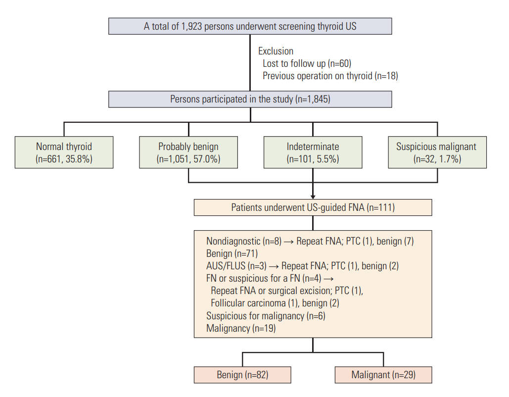
Fig. 1. Flow diagram of the study. US, ultrasonography; FNA, fine-needle aspiration; PTC, papillary thyroid carcinoma; AUS/FLUS, atypia of undetermined significance or follicular lesions of undetermined significance; FN, follicular neoplasm.

Fig. 2. A 54-year-old man with a 1.2 cm suspicious malignant nodule in the left thyroid gland. (A, B) Axial and longitudinal ultrasonography images show a hypoechoic nodule with a taller-than-wide axis, ill-defined margin, and multiple hyperechoic foci suggesting micro/macrocalcifications (arrows). Upon ultrasonography-guided fine needle aspiration and pathological evaluation, the nodule was revealed to be a papillary thyroid carcinoma. The patient underwent total thyroidectomy, and the final diagnosis was papillary thyroid carcinoma.
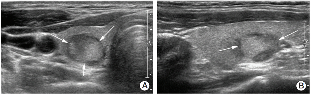
Fig. 3. A 40-year-old man with a 1.3-cm indeterminate nodule in the right thyroid gland. (A, B) Axial and longitudinal ultrasonography (US) images show an oval solid isoechoic nodule with wider-than-tall axis (arrows). Upon US-guided fine needle aspiration and pathological evaluation, the nodule was found to be suspicious for follicular neoplasm. The patient underwent total thyroidectomy, and the final diagnosis was minimally invasive follicular carcinoma (R-minor-2).
Cited by 1 articles
-
Changes in the Diagnostic Efficiency of Thyroid Fine-Needle Aspiration Biopsy during the Era of Increased Thyroid Cancer Screening in Korea
Young Ki Lee, Kyeong Hye Park, Young Duk Song, Taemi Youk, Joo Young Nam, Sun Ok Song, Dong Yeob Shin, Eun Jig Lee
Cancer Res Treat. 2019;51(4):1430-1436. doi: 10.4143/crt.2018.534.
Reference
-
References
1. Vander JB, Gaston EA, Dawber TR. The significance of nontoxic thyroid nodules. Final report of a 15-year study of the incidence of thyroid malignancy. Ann Intern Med. 1968; 69:537–40.2. Ezzat S, Sarti DA, Cain DR, Braunstein GD. Thyroid incidentalomas: prevalencee by palpation and ultrasonography. Arch Intern Med. 1994; 154:1838–40.3. Mortensen JD, Woolner LB, Bennett WA. Gross and microscopic findings in clinically normal thyroid glands. J Clin Endocrinol Metab. 1955; 15:1270–80.
Article4. Watters DA, Ahuja AT, Evans RM, Chick W, King WW, Metreweli C, et al. Role of ultrasound in the management of thyroid nodules. Am J Surg. 1992; 164:654–7.
Article5. Steele SR, Martin MJ, Mullenix PS, Azarow KS, Andersen CA. The significance of incidental thyroid abnormalities identified during carotid duplex ultrasonography. Arch Surg. 2005; 140:981–5.
Article6. Frates MC, Benson CB, Charboneau JW, Cibas ES, Clark OH, Coleman BG, et al. Management of thyroid nodules detected at US: Society of Radiologists in Ultrasound consensus conference statement. Radiology. 2005; 237:794–800.
Article7. Kilfoy BA, Zheng T, Holford TR, Han X, Ward MH, Sjodin A, et al. International patterns and trends in thyroid cancer incidence, 1973-2002. Cancer Causes Control. 2009; 20:525–31.
Article8. Davies L, Welch HG. Increasing incidence of thyroid cancer in the United States, 1973-2002. JAMA. 2006; 295:2164–7.
Article9. Ahn HS, Kim HJ, Welch HG. Korea's thyroid-cancer “epidemic”: screening and overdiagnosis. N Engl J Med. 2014; 371:1765–7.10. Chen AY, Jemal A, Ward EM. Increasing incidence of differentiated thyroid cancer in the United States, 1988-2005. Cancer. 2009; 115:3801–7.
Article11. Morris LG, Myssiorek D. Improved detection does not fully explain the rising incidence of well-differentiated thyroid cancer: a population-based analysis. Am J Surg. 2010; 200:454–61.
Article12. Wartofsky L. Increasing world incidence of thyroid cancer: increased detection or higher radiation exposure? Hormones (Athens). 2010; 9:103–8.
Article13. Jung KW, Won YJ, Kong HJ, Oh CM, Cho H, Lee DH, et al. Cancer statistics in Korea: incidence, mortality, survival, and prevalence in 2012. Cancer Res Treat. 2015; 47:127–41.
Article14. Yi KH, Kim SY, Kim DH, Kim SW, Na DG, Lee YJ, et al. The Korean guideline for thyroid cancer screening. J Korean Med Assoc. 2015; 58:302–12.
Article15. Kim JY, Lee CH, Kim SY, Jeon WK, Kang JH, An SK, et al. Radiologic and pathologic findings of nonpalpable thyroid carcinomas detected by ultrasonography in a medical screening center. J Ultrasound Med. 2008; 27:215–23.
Article16. Choi YJ, Park YL, Koh JH. Prevalence of thyroid cancer at a medical screening center: pathological features of screendetected thyroid carcinomas. Yonsei Med J. 2008; 49:748–56.
Article17. Liel Y. Screening without evidence of efficacy: thyroid ultrasonography is another example. BMJ. 2004; 328:521.18. Moon WJ, Baek JH, Jung SL, Kim DW, Kim EK, Kim JY, et al. Ultrasonography and the ultrasound-based management of thyroid nodules: consensus statement and recommendations. Korean J Radiol. 2011; 12:1–14.
Article19. Cibas ES, Ali SZ; NCI Thyroid FNA State of the Science Conference. The Bethesda System For Reporting Thyroid Cytopathology. Am J Clin Pathol. 2009; 132:658–65.
Article20. Kang HW, No JH, Chung JH, Min YK, Lee MS, Lee MK, et al. Prevalence, clinical and ultrasonographic characteristics of thyroid incidentalomas. Thyroid. 2004; 14:29–33.
Article21. Haugen BR, Alexander EK, Bible KC, Doherty GM, Mandel SJ, Nikiforov YE, et al. 2015 American Thyroid Association management guidelines for adult patients with thyroid nodules and differentiated thyroid cancer: The American Thyroid Association Guidelines Task Force on Thyroid Nodules and Differentiated Thyroid Cancer. Thyroid. 2016; 26:1–133.
Article22. Russ G, Leboulleux S, Leenhardt L, Hegedus L. Thyroid incidentalomas: epidemiology, risk stratification with ultrasound and workup. Eur Thyroid J. 2014; 3:154–63.
Article23. Nam-Goong IS, Kim HY, Gong G, Lee HK, Hong SJ, Kim WB, et al. Ultrasonography-guided fine-needle aspiration of thyroid incidentaloma: correlation with pathological findings. Clin Endocrinol (Oxf). 2004; 60:21–8.
Article24. Kim DW, Eun CK, In HS, Kim MH, Jung SJ, Bae SK. Sonographic differentiation of asymptomatic diffuse thyroid disease from normal thyroid: a prospective study. AJNR Am J Neuroradiol. 2010; 31:1956–60.
Article25. Berg WA, Blume JD, Cormack JB, Mendelson EB, Lehrer D, Bohm-Velez M, et al. Combined screening with ultrasound and mammography vs mammography alone in women at elevated risk of breast cancer. JAMA. 2008; 299:2151–63.
Article26. Moon HJ, Jung I, Park SJ, Kim MJ, Youk JH, Kim EK. Comparison of cancer yields and diagnostic performance of screening mammography vs. supplemental screening ultrasound in 4394 women with average risk for breast cancer. Ultraschall Med. 2015; 36:255–63.27. Solares CA, Penalonzo MA, Xu M, Orellana E. Occult papillary thyroid carcinoma in postmortem species: prevalence at autopsy. Am J Otolaryngol. 2005; 26:87–90.
Article28. Neuhold N, Kaiser H, Kaserer K. Latent carcinoma of the thyroid in Austria: a systematic autopsy study. Endocr Pathol. 2001; 12:23–31.
Article29. Lee J, Rhee Y, Lee S, Ahn CW, Cha BS, Kim KR, et al. Frequent, aggressive behaviors of thyroid microcarcinomas in korean patients. Endocr J. 2006; 53:627–32.
Article30. Ito Y, Uruno T, Nakano K, Takamura Y, Miya A, Kobayashi K, et al. An observation trial without surgical treatment in patients with papillary microcarcinoma of the thyroid. Thyroid. 2003; 13:381–7.
Article
- Full Text Links
-
- Actions
-
Cited
- CITED
-
- Close
- Share
-
- Similar articles
-
- Thyroid cancer screening
- Screening of Thyroid Cancer and Management of Thyroid Incidentaloma
- Value of Ultrasonographic Mass Screening for Thyroid Carcinoma in Patients Undergoing a Breast Ultrasonography
- A Refutation against Unfounded Reports on Thyroid Cancer
- Evaluation of Mass Screening for Thyroid Cancer
