Full mouth rehabilitation of severely worn dentition with implants and removable partial dentures
- Affiliations
-
- 1Department of Prosthodontics, Seoul St. Mary's Hospital, College of Medicine, The Catholic University of Korea, Seoul, Repulic of Korea. lsuyoung@daum.net
- KMID: 2402990
- DOI: http://doi.org/10.4047/jkap.2018.56.1.70
Abstract
- Excessive tooth wear can lead to decrease in occlusal vertical dimension and can cause pathological changes in the oral environment and masticatory system. When recovering occlusal vertical dimension and occlusion, accurate diagnosis and analysis are essential. This clinical case describes a 75-year-old woman with severely worn dentition due to loss of the posterior support. Full mouth rehabilitation with occlusal vertical dimension increment was planned. Clinical and radiographic examinations, occlusal vertical dimension evaluation, and diagnostic wax-up were performed and patient adaptability was evaluated using provisional restorations. As for definitive restoration, considering economic condition of the patient, removable partial denture was fabricated and solitary implants were placed in the mandibular left and right posterior region to increase support and retention of the removable partial denture. During one year of follow-up, functional and esthetic outcomes were observed satisfactory.
Keyword
MeSH Terms
Figure
-

Fig. 1 Intraoral view of the patient before treatment. Severely worn mandibular natural teeth occluding maxillary PFM prosthesis were observed. (A) Maxillary occlusal view, (B) Frontal view, (C) Mandibular occlusal view.
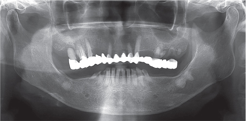
Fig. 2 Initial panoramic radiograph before treatment. Wears on occlusal surface of the mandibular teeth and severe secondary caries with peri-apical lesions on the maxillary posterior teeth were observed.
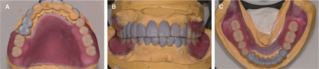
Fig. 3 Diagnostic wax up at increased OVD. (A) Maxillary occlusal view, (B) Frontal view, (C) Mandibular occlusal view.

Fig. 4 Provisional restoration. (A) Maxillary occlusal view, (B) Frontal view, (C) Mandibular occlusal view.

Fig. 5 Cone beam computed tomography image for implant placement. (A) Mandibular right second molar, (B) Mandibular left second molar.
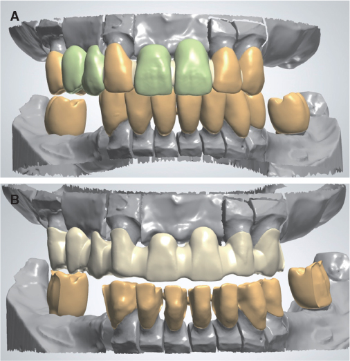
Fig. 6 Computer-aided design for porcelain fused metal framework fabrication using laser sintering technique. (A) Full contour design, (B) Cut-back.
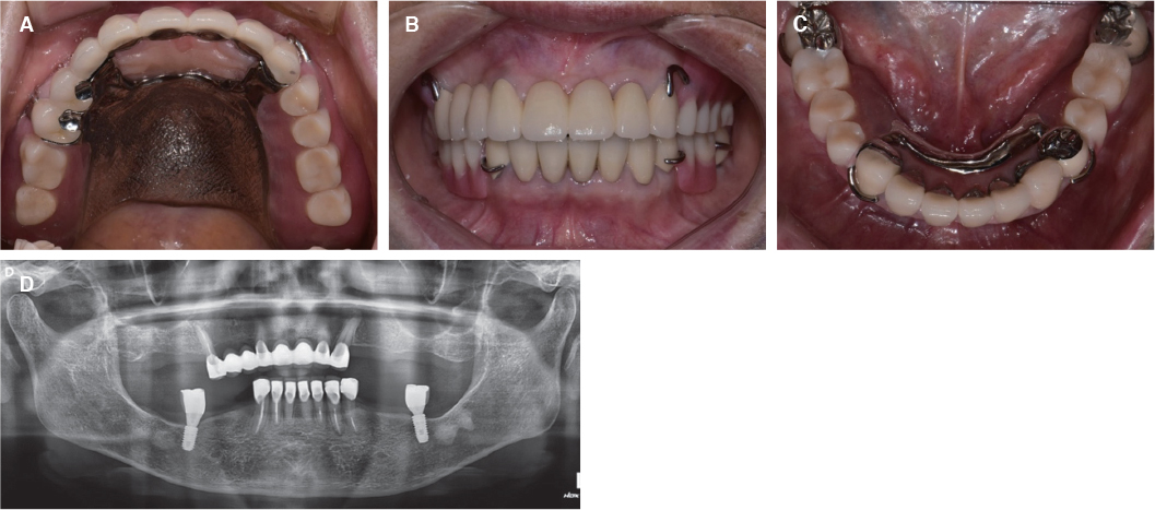
Fig. 7 Definitive prostheses. (A) Maxillary occlusal view, (B) Frontal view, (C) Mandibular occlusal view, (D) Panoramic radiograph.
Reference
-
1. Hattab FN, Yassin OM. Etiology and diagnosis of tooth wear: a literature review and presentation of selected cases. Int J Prosthodont. 2000; 13:101–107.2. Smith BG, Knight JK. A comparison of patterns of tooth wear with aetiological factors. Br Dent J. 1984; 157:16–19.
Article3. Smith BG, Knight JK. An index for measuring the wear of teeth. Br Dent J. 1984; 156:435–438.
Article4. Dawson PE. Functional occlusion: from TMJ to smile design. St. Louis; Mo: Mosby;2007. p. 430–452.5. Jahangiri L, Jang S. Onlay partial denture technique for assessment of adequate occlusal vertical dimension: a clinical report. J Prosthet Dent. 2002; 87:1–4.
Article6. Muts EJ, van Pelt H, Edelhoff D, Krejci I, Cune M. Tooth wear: a systematic review of treatment options. J Prosthet Dent. 2014; 112:752–759.7. Turner KA, Missirlian DM. Restoration of the extremely worn dentition. J Prosthet Dent. 1984; 52:467–474.
Article8. Ibbetson RJ, Setchell DJ. Treatment of the worn dentition: 2. Dent Update. 1989; 16:300–302. 305–307.9. Grippo JO, Simring M, Schreiner S. Attrition, abrasion, corrosion and abfraction revisited: a new perspective on tooth surface lesions. J Am Dent Assoc. 2004; 135:1109–1118.10. Verrett RG. Analyzing the etiology of an extremely worn dentition. J Prosthodont. 2001; 10:224–233.
Article11. Abduo J, Lyons K. Clinical considerations for increasing occlusal vertical dimension: a review. Aust Dent J. 2012; 57:2–10.
Article12. Tang Y, Loh HT, Wong YS, Fuh JYH, Lu L, Wang X. Direct laser sintering of a copper-based alloy for creating three-dimensional metal parts. J Mat Proc Tech. 2003; 140:368–372.
Article13. Akova T, Ucar Y, Tukay A, Balkaya MC, Brantley WA. Comparison of the bond strength of laser-sintered and cast base metal dental alloys to porcelain. Dent Mater. 2008; 24:1400–1404.
Article14. Keltjens HM, Kayser AF, Hertel R, Battistuzzi PG. Distal extension removable partial dentures supported by implants and residual teeth: considerations and case reports. Int J Oral Maxillofac Implants. 1993; 8:208–213.15. Kuzmanovic DV, Payne AG, Purton DG. Distal implants to modify the Kennedy classification of a removable partial denture: a clinical report. J Prosthet Dent. 2004; 92:8–11.
Article16. Schneid TR, Mattie PA. Implant-assisted removable partial dentures. In : Phoenix RD, Cagna DR, DeFreest CF, editors. Stewart's clinical removable partial prosthodontics. 4th ed. Chicago: Quintessence Publishing Company;2008. p. 259–277.17. Jang Y, Emtiaz S, Tarnow DP. Single implant-supported crown used as an abutment for a removable cast partial denture: a case report. Implant Dent. 1998; 7:199–204.
Article18. Pellecchia M, Pellecchia R, Emtiaz S. Distal extension mandibular removable partial denture connected to an anterior fixed implant-supported prosthesis: a clinical report. J Prosthet Dent. 2000; 83:607–612.
Article19. Starr NL. The distal extension case: an alternative restorative design for implant prosthetics. Int J Periodontics Restorative Dent. 2001; 21:61–67.20. Chronopoulos V, Sarafianou A, Kourtis S. The use of dental implants in combination with removable partial dentures: a case report. J Esthet Restor Dent. 2008; 20:355–364.
Article
- Full Text Links
-
- Actions
-
Cited
- CITED
-
- Close
- Share
-
- Similar articles
-
- Full mouth rehabilitation with a few remaining teeth and implants for a patient with chronic periodontitis: a case report
- Three-year follow-up of full mouth rehabilitation with anterior implant surveyed bridges and distal extension removable partial denture
- Full mouth rehabilitation using removable partial denture in patient with loss of vertical dimension due to worn dentition
- Computer-aided design and manufacturing-based full mouth rehabilitation for a patient with excessive attrition and restricted vertical dimension: A case report
- Full mouth rehabilitation with extremely worn dentition
