Local Hyperthermia Affects Murine Contact Hypersensitivity around Elicitation Phase
- Affiliations
-
- 1Department of Dermatology, No. 1 Hospital of China Medical University, Shenyang, China. jlwuyan@126.com, gaobarry@hotmail.com
- KMID: 2399769
- DOI: http://doi.org/10.5021/ad.2018.30.1.107
Abstract
- No abstract available.
MeSH Terms
Figure
-

Fig. 1 (A) Comparison of the difference values at different temperatures. The difference values of 39℃, 41℃, or 43℃ were higher than that of 37℃, respectively. The difference value of 43℃ was higher than those of 39℃ or 41℃, respectively. No significant difference was seen in concurrent-heated group and post-heated group among different temperatures. *Represented significant difference comparing to 37℃ (p<0.05), †represented significant difference comparing to 39℃ (p<0.05), ‡represented significant difference comparing to 41℃ (p<0.05). (B) Comparison of the difference values of different groups. No significant difference was seen among the three group at 37℃ or 43℃. The difference value of post-heated group were significantly higher than that of pre-heated group at 39℃ or 41℃. Represented significant difference comparing to pre-heated group (p<0.05).
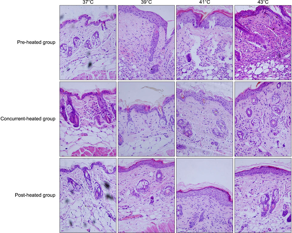
Fig. 2 Histological examination (H&E, ×40). The histological manifestations of pre-heated group, concurrent-heated group and post-heated group were similar. The manifestations of control and 37℃ were almost the same. The intracellular and/or intercellular edema of epidermis was slight at 39℃, and moderate at 41℃ and 43℃. Increment of lymphocytes were seen in both epidermis and dermis, at 39℃, 41℃, and 43℃.
Reference
-
1. Christensen AD, Haase C. Immunological mechanisms of contact hypersensitivity in mice. APMIS. 2012; 120:1–27.
Article2. Timares L, Katiyar SK, Elmets CA. DNA damage, apoptosis and langerhans cells--activators of UV-induced immune tolerance. Photochem Photobiol. 2008; 84:422–436.
Article3. Lesiak A, Norval M, Sysa-Jedrzejowska A, Wozniacka A, Kobos J, Omulecka A, et al. Elicitation of contact hypersensitivity after repeated suberythemal exposures of humans to solar simulated radiation: number of epidermal Langerhans cells. Contact Dermatitis. 2007; 57:224–229.
Article4. Hall JM, Witter AR, Racine RR, Berg RE, Podawiltz A, Jones H, et al. Chronic psychological stress suppresses contact hypersensitivity: potential roles of dysregulated cell trafficking and decreased IFN-γ production. Brain Behav Immun. 2014; 36:156–164.
Article5. Danno K, Sugie N. Effects of near-infrared radiation on the epidermal proliferation and cutaneous immune function in mice. Photodermatol Photoimmunol Photomed. 1996; 12:233–236.
Article6. Ostberg JR, Gellin C, Patel R, Repasky EA. Regulatory potential of fever-range whole body hyperthermia on Langerhans cells and lymphocytes in an antigen-dependent cellular immune response. J Immunol. 2001; 167:2666–2670.
Article7. Zhang L, Wang YR, Hong YX, Xu YQ, Zhang L, Li XD, et al. Temporal effect of local hyperthermia on murine contact hypersensitivity. Chin Med J (Engl). 2013; 126:1555–1559.8. Koyama Y, Nagao S, Ohashi K, Takahashi H, Marunouchi T. Effect of systemic and topical application of testosterone propionate on the density of epidermal Langerhans cells in the mouse. J Invest Dermatol. 1989; 92:86–90.
Article9. Dudeck J, Ghouse SM, Lehmann CH, Hoppe A, Schubert N, Nedospasov SA, et al. Mast-cell-derived TNF amplifies CD8(+) dendritic cell functionality and CD8(+) T cell priming. Cell Rep. 2015; 13:399–411.
Article10. Honda T, Matsuoka T, Ueta M, Kabashima K, Miyachi Y, Narumiya S. Prostaglandin E(2)-EP(3) signaling suppresses skin inflammation in murine contact hypersensitivity. J Allergy Clin Immunol. 2009; 124:809–818.e2.
Article
- Full Text Links
-
- Actions
-
Cited
- CITED
-
- Close
- Share
-
- Similar articles
-
- The Effect of Topical Tacrolimus in the Murine Contact Hypersensitivity and Dermatitis of Repeated Applications Induced by Diphenylcyclopropenone
- Hyperthermia Depletes Epidermal Langerhans Cells and Modulates Contact Hypersensitivity Reaction in Mice
- Effects of UVB on the Induction and Elicitation of DNCB Contact Sensitivity in Guinea Pigs
- Effect of Cimetidine on Contact Sensitivity Reaction in Guinea Pigs
- High Doses of UVA Suppress Contact Hypersensitivity
