Asymptomatic Hyperpigmentation without Preceding Inflammation as a Clinical Feature of Citrus Fruits-Induced Phytophotodermatitis
- Affiliations
-
- 1Department of Dermatology, Gangnam Severance Hospital, Cutaneous Biology Research Institute, Yonsei University College of Medicine, Seoul, Korea.
- 2Department of Dermatology and Cutaneous Biology Research Institute, Severance Hospital, Yonsei University College of Medicine, Seoul, Korea. oddung93@yuhs.ac
- KMID: 2399758
- DOI: http://doi.org/10.5021/ad.2018.30.1.75
Abstract
- Phytophotodermatitis is a condition that occurs by contact with plants containing phototoxic agents such as furocoumarins and psoralens with subsequent ultraviolet exposure. Phytophotodermatitis typically presents as sharply defined erythematous patches with occasional blistering, sometimes accompanied with pain or itching sensation. In some cases, however, sudden appearance of asymptomatic hyperpigmentation can be the only clinical finding of phytophotodermatitis. Here, we present two patients with sudden development of asymptomatic pigmentation on their hand without preceding inflammation by the contact with citrus fruits containing photosensitizers and subsequent exposure to strong sunlight. As like these patients, phytophotodermatitis can present with only pigmentation without noticeable inflammation especially in dark skinned people. In such cases, physician can sometimes have difficulty in diagnosis of phytophotodermatitis. Therefore, it is important to consider the possibility of phytophotodermatitis through careful history taking, especially in patients who have abruptly developed well-defined hyperpigmentation on sun-exposed areas, to avoid unnecessary test and treatment.
Keyword
MeSH Terms
Figure
-
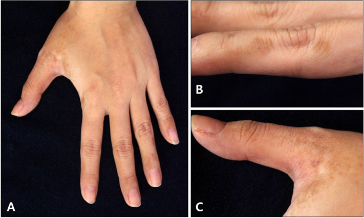
Fig. 1 (A) Multiple brown pigmented patches on the left hand. (B) On the lateral sides of fingers, pigmentations showed irregular shape with rather sharp borders. (C) Some pigmentation showed reticulated pattern.
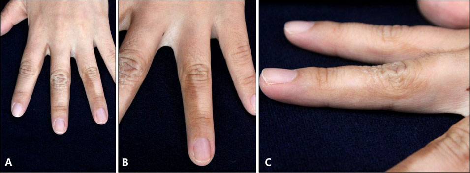
Fig. 2 (A) Multiple brown pigmented patches on the left hand. (B) Homogeneous brown colored pigmentations with rather straight borders were observed on the lateral sides and the dorsum of fingers. (C) Some pigmentations showed overlying scales.
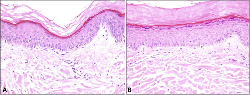
Fig. 3 (A) Skin biopsy specimen from case 1. (B) Skin biopsy specimen from case 2. Both skin tissues showed slightly increased number of melanocytes and basal hyperpigmentation (H&E; A, B: ×200).
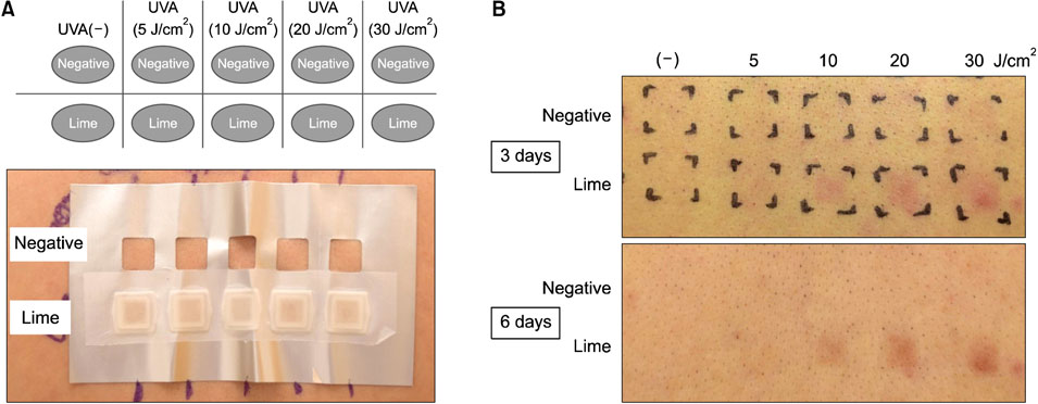
Fig. 4 (A) Schematic design of photoprovocation test and back skin of a healthy adult male. Skin was split into untreated (negative) and lime extract applied area. The 5, 10, 20, and 30 J/cm2 of ultraviolet A (UVA) were irradiated on the back. (B) Three days and six days after UVA irradiation. Brown colored, homogeneous pigmented macules are observed.
Reference
-
1. dos Reis VM. Dermatosis due to plants (phytodermatosis). An Bras Dermatol. 2010; 85:479–489.2. Cestari TF, Dantas LP, Boza JC. Acquired hyperpigmentations. An Bras Dermatol. 2014; 89:11–25.
Article3. Galvañ-Pérez Del, Linares-Barrios M, Galvañ-Pozo JI Jr. Acropigmentation of the dorsum of the hands from preparing mojitos: a lime-induced phytophotodermatosis. Actas Dermosifiliogr. 2016; 107:253–255.
Article4. Goskowicz MO, Friedlander SF, Eichenfield LF. Endemic “lime” disease: phytophotodermatitis in San Diego County. Pediatrics. 1994; 93:828–830.
Article
- Full Text Links
-
- Actions
-
Cited
- CITED
-
- Close
- Share
-
- Similar articles
-
- Phytophotodermatitis due to a Citrus-Based Hand Sanitizer: A Case Report
- Citrus Fruit Intake and Breast Cancer Risk: A Quantitative Systematic Review
- Citrus Fruits Intake and Prostate Cancer Risk: A Quantitative Systematic Review
- Clinical Course of Bimatoprost-induced Periocular Skin Hyperpigmentation after Stopping Bimatoprost Treatment
- A Case of Progressive Cribriform and Zosteriform Hyperpigmentation Associated with Becker's Nevus and Melanonychia
